. 2006 Jun 15;573(Pt 3):711-21.
doi: 10.1113/jphysiol.2006.109447. Epub 2006 Apr 13.

Activity-dependent synaptic plasticity in the supraoptic nucleus of the rat hypothalamus

Affiliations

Activity-dependent synaptic plasticity in the supraoptic nucleus of the rat hypothalamus

Aude Panatier et al. J Physiol. .

Abstract

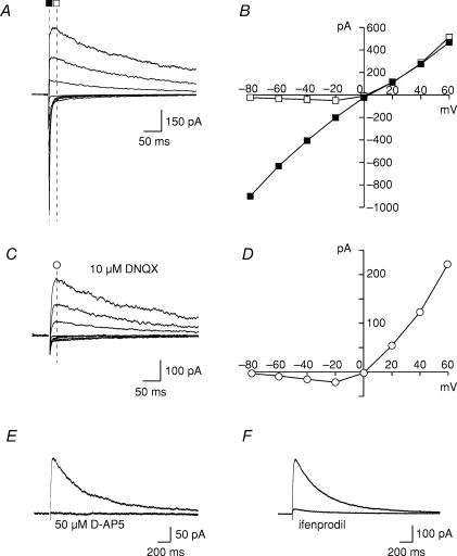
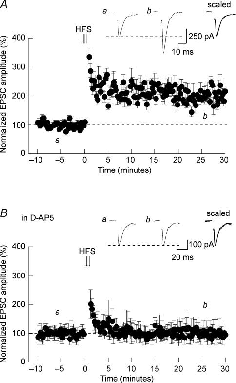
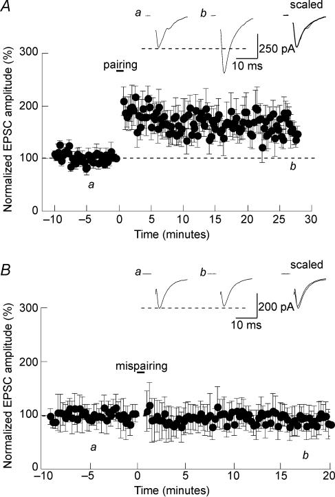
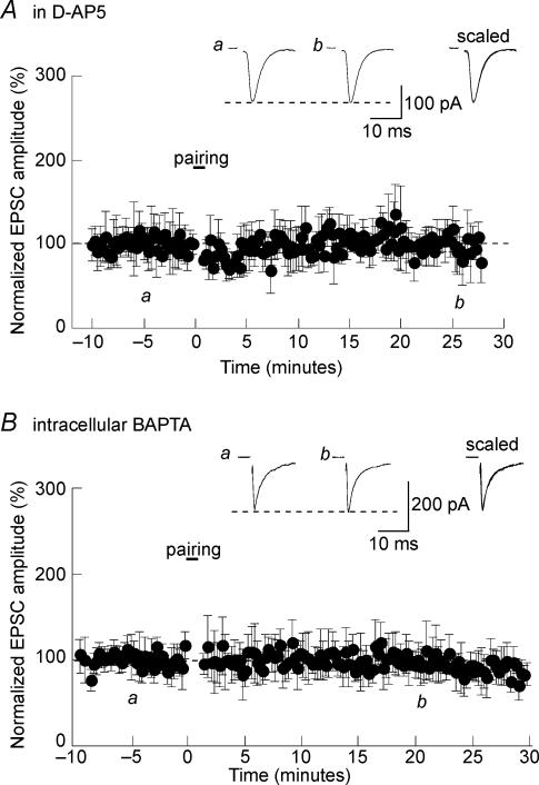
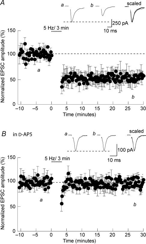
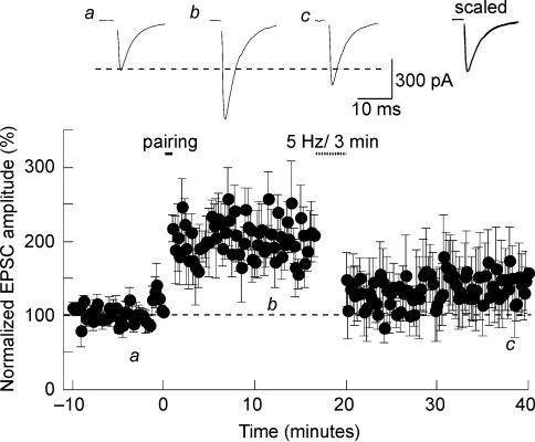
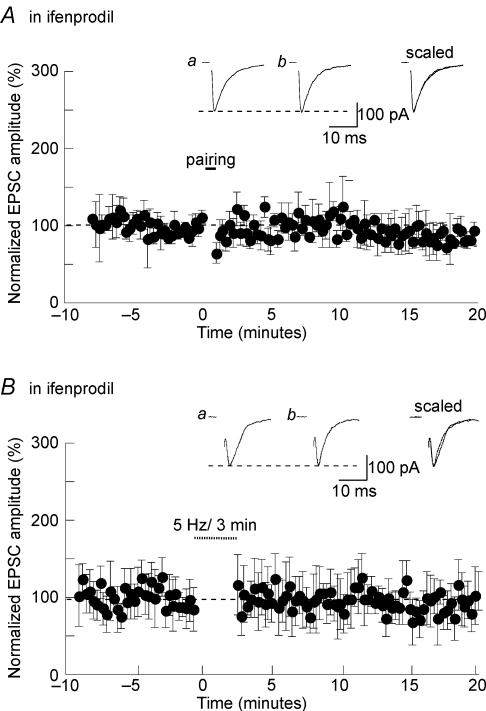
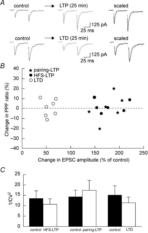
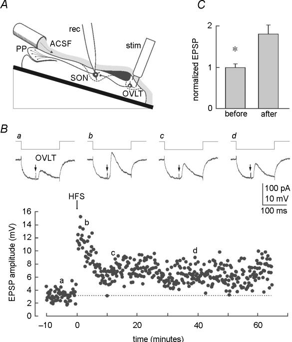
Activity-dependent long-term synaptic changes were investigated at glutamatergic synapses in the supraoptic nucleus (SON) of the rat hypothalamus. In acute hypothalamic slices, high frequency stimulation (HFS) of afferent fibres caused long-term potentiation (LTP) of the amplitude of AMPA receptor-mediated excitatory postsynaptic currents (EPSCs) recorded with the whole-cell patch-clamp technique. LTP was also obtained in response to membrane depolarization paired with mild afferent stimulation. On the other hand, stimulating the inputs at 5 Hz for 3 min at resting membrane potential caused long-term depression (LTD) of excitatory transmission in the SON. These forms of synaptic plasticity required the activation of NMDA receptors since they were abolished in the presence of D-AP5 or ifenprodil, two selective blockers of these receptors. Analysis of paired-pulse facilitation and trial-to-trial variability indicated that LTP and LTD were not associated with changes in the probability of transmitter release, thereby suggesting that the locus of expression of these phenomena was postsynaptic. Using sharp microelectrode recordings in a hypothalamic explant preparation, we found that HFS also generates LTP at functionally defined glutamatergic synapses formed between the organum vasculosum lamina terminalis and SON neurons. Taken together, our findings indicate that glutamatergic synapses in the SON exhibit activity-dependent long-term synaptic changes similar to those prevailing in other brain areas. Such forms of plasticity could play an important role in the context of physiological responses, like dehydration or lactation, where the activity of presynaptic glutamatergic neurons is strongly increased.


Figures

Figure 1
Figure 1. Properties of synaptic NMDAR-mediated responses
A, evoked EPSCs obtained from a representative SON neuron at different membrane potentials (from −80 to +60 mV). Traces are averages of 5–10 consecutive sweeps. B, current–voltage (IV) relationships obtained from the traces in A, 5 ms (▪) and 40 ms (□) after stimulation. Note the rectification when the current was measured later on the trace. C and D, evoked EPSCs and corresponding IV curve obtained in the presence of DNQX. Traces are averages of 5–10 consecutive sweeps. E and F, representative examples of the inhibitory effects of 50 μm D-AP5 and 10 μm ifenprodil on evoked EPSCs recorded in the presence of DNQX. Traces are averages of 5–10 consecutive sweeps.
Figure 2
Figure 2. HFS-induced LTP in the SON
A, in rat hypothalamic slices, a high frequency stimulation (arrows) induced LTP of evoked EPSCs. The insets are averaged sample records of 10 sweeps taken at the times indicated from a typical experiment. These traces were superimposed and scaled. B, the same HFS protocol did not induced LTP when given in the presence of 50 μmd-AP5.
Figure 3
Figure 3. Pairing-induced LTP in the SON
A, in rat SON neurons, pairing afferent stimulation (2 Hz) with membrane depolarization (0 mV) for 45 s caused LTP of evoked EPSCs. B, stimulating the afferent inputs at 2 Hz without depolarizing the neurons (mispairing) failed to induce LTP.
Figure 4
Figure 4. Involvement of NMDARs in LTP induction in the SON
A, in the presence of 50 μmd-AP5, pairing afferent stimulation with membrane depolarization failed to induce LTP. B, pairing-LTP was prevented when BAPTA was included in the patch pipette to buffer intracellular Ca2+.
Figure 5
Figure 5. Induction of NMDAR-LTD in the SON
A, in rat hypothalamic slices, stimulating the afferent fibres at 5 Hz for 3 min in current clamp mode at resting membrane potential induced LTD of evoked EPSCs. B, the same protocol did not induced LTD when given in the presence of 50 μmd-AP5.
Figure 6
Figure 6. Depotentiation in the SON
Synaptic strength can be sequentially increased and decreased at a single set of synapses. Synapses were first potentiated by a pairing protocol and then depotentiated 15 min later with a 5-Hz-3 min train of stimulation.
Figure 7
Figure 7. Involvement of NR2B-containing NMDARs in LTP and LTD in the SON
Pairing-LTP (A) and LTD (B) were prevented when NR2B-containing NMDA receptors were blocked with 10 μm ifenprodil.
Figure 8
Figure 8. Postsynaptic locus of expression of LTP and LTD in the SON
A, representative examples where a pair of stimulations was applied at 50 ms interval before (control) and after induction of HFS-LTP (top) and LTD (bottom). Traces, which are averages of 5–10 consecutive sweeps, were superimposed and scaled to the amplitude of the first EPSC obtained under control conditions (right panel). Note that LTP and LTD did not affect the paired-pulse facilitation. B, plot summarizing the change in PPF (y-axis) as a function of the change in EPSC amplitude (x-axis) associated with HFS-LTP (•), pairing LTP (⋆) and LTD (○). C, summary bar graph of 1/CV2 values measured before (filled bars) and after (open bars) the induction of HFS-LTP, pairing LTP and LTD. Note that 1/CV2 was not affected by any of these long-term changes.
Figure 9
Figure 9. LTP of glutamatergic synapses in the OVLT-SON pathway
A, schematic diagram providing a side view of the experimental preparation. The tube delivering artificial cerebrospinal fluid (ACSF) is positioned just rostral and lateral to the posterior pituitary (PP), while stimulating (stim) and recording (rec) electrodes are placed in the OVLT and SON, respectively. B, representative intracellular recording experiment made in one SON neuron. Upper panels (ad) show EPSPs evoked in an SON neuron by stimulation of the OVLT (arrows). Each trace is an average of 4 consecutive sweeps taken at various times before and after HFS (see corresponding letters in lower graph). Note that each synaptic response is elicited during the steady-state phase of the electrotonic response to a hyperpolarizing current pulse delivered to monitor input resistance. The lower graph plots the absolute amplitude of EPSPs evoked in the same cell during a representative experiment. Note that input resistance is unaffected while a sustained increase in EPSP amplitude can be observed for more than 1 h following HFS. C, bar graph showing the normalized amplitude of OVLT-mediated EPSPs recorded in 7 SON neurons before and > 30 min following HFS.

Similar articles

Cited by

References

    1. Belin V, Moos F. Paired recordings from supraoptic and paraventricular oxytocin cells in suckled rats: recruitment and synchronization. J Physiol. 1986;377:369–390. - PMC - PubMed
    1. Csaki A, Kocsis K, Kiss J, Halasz B. Localization of putative glutamatergic/aspartatergic neurons projecting to the supraoptic nucleus area of the rat hypothalamus. Eur J Neurosci. 2002;16:55–68. - PubMed
    1. Cummings JA, Mulkey RM, Nicoll RA, Malenka RC. Ca2+ signaling requirements for long-term depression in the hippocampus. Neuron. 1996;16:825–833. - PubMed
    1. De Kock CP, Burnashev N, Lodder JC, Mansvelder HD, Brussaard AB. NMDA receptors induce somatodendritic secretion in hypothalamic neurones of lactating female rats. J Physiol. 2004;561:53–64. - PMC - PubMed
    1. Gentles SJ. Montreal, Canada: McGill University; 1987. Patterned afferent activity and synaptic plasticity in the magnocellular neurosecretory system. MSc Thesis.

Publication types

MeSH terms

LinkOut - more resources