Comparative Study
. 2006 Jun 15;573(Pt 3):753-63.
doi: 10.1113/jphysiol.2006.108613. Epub 2006 Apr 13.

Direct excitation of deep dorsal horn neurones in the rat spinal cord by the activation of postsynaptic P2X receptors

Affiliations
Comparative Study

Direct excitation of deep dorsal horn neurones in the rat spinal cord by the activation of postsynaptic P2X receptors

Hiroaki Shiokawa et al. J Physiol. .

Abstract

ATP mediates somatosensory transmission in the spinal cord through the activation of P2X receptors. Nonetheless, the functional significance of postsynaptic P2X receptors in spinal deep dorsal horn neurones is still not yet well understood. Using the whole-cell patch-clamp technique, we investigated whether the activation of postsynaptic P2X receptors can modulate the synaptic transmission in lamina V neurones of postnatal day (P) 9-12 spinal cord slices. At a holding potential of -70 mV, ATPgammaS (100 microm), a nonhydrolysable ATP analogue, generated an inward current, which was resistant to tetrodotoxin (1 microm) in 61% of the lamina V neurones. The ATPgammaS-induced inward current was accompanied by a significant increase in the frequency of glutamatergic miniature excitatory postsynaptic currents (mEPSCs) in the majority of lamina V neurones. The ATPgammaS-induced inward current was not reproduced by P2Y receptor agonists, UTP (100 microm), UDP (100 microm), and 2-methylthio ADP (100 microm), and it was also not affected by the addition of guanosine-5'-O-(2-thiodiphosphate) (GDPbetaS) into the pipette solution, thus suggesting that ionotropic P2X receptors were activated by ATPgammaS instead of metabotropic P2Y receptors. On the other hand, alpha,beta-methylene ATP (100 microm) did not change any membrane current, but instead increased the mEPSC frequency in the majority of lamina V neurones. The ATPgammaS-induced inward current was suppressed by pyridoxalphosphate-6-azophenyl-2',4'-disulphonic acid (PPADS) (10 microm), but not by trinitrophenyl-ATP (TNP-ATP) (1 microm). Furthermore, we found that ATPgammaS (100 microm) produced a clear inward current which was observed in all lamina V neurones over P16 spinal cord slices, in contrast to P9-12. These results indicate that distinct subtypes of P2X receptors were functionally expressed at the post- and presynaptic sites in lamina V neurones, both of which may contribute to the hyperexcitability of lamina V in a different manner. In addition, the data relating to the developmental increase in the functional P2X receptors suggest that purinergic signalling may thus be more common in somatosensory transmission with maturation.


Figures

Figure 1
Figure 1. ATPγS excites lamina V neurones in spinal cord slices
A, a recorded lamina V neurone identified with an intracellular injection with neurobiotin. Scale bar is 100 μm. B, all lamina V neurones examined displayed tonic firing in response to depolarizing current injection of 143.6 pA (upper trace), 93.6 pA (middle trace), or 43.6 pA (lower trace) in current-clamp mode. C, in voltage-clamp mode, ATPγS (100 μm) produced a clear inward current at a holding potential of −70 mV (upper trace). In the presence of CNQX (20 μm) and APV (50 μm), ATPγS (100 μm) still induced an inward current without a significant decrease in amplitude in the same lamina V neurone (lower trace).
Figure 2
Figure 2. Post- or presynaptic effect of ATPγS in lamina V neurones
A, bath application of ATPγS (100 μm) for 1 min induced a clear inward current associated with a marked increase in the sEPSC frequency in a P9–12 lamina V neurone. The ATPγS-induced inward current was observed in approximately 60% of lamina V neurones examined. B, a summary of relative sEPSC frequency before (control) and during application of ATPγS in lamina V neurones which exhibited the ATPγS-induced inward current (*P < 0.05). C, ATPγS (100 μm) did not change any membrane current, but largely increased the sEPSC frequency in a P9–P12 lamina V neurone. D, a summary of the relative sEPSC frequency before (control) and during the application of ATPγS in lamina V neurones which did not exhibit any ATPγS-induced inward current (*P < 0.05). Error bars s.e.m.
Figure 3
Figure 3. ATPγS repeatedly induces an inward current in lamina V neurones
A, when ATPγS was repeatedly applied at 10 min intervals, it produced a similar inward current (upper trace, the first application of ATPγS; lower trace, the second application of ATPγS). B, in the presence of TTX (1 μm), ATPγS induced an inward current without any decrease in the amplitude (upper trace, in the absence of TTX; lower trace, in the presence of TTX).
Figure 4
Figure 4. Dose and voltage dependency of the ATPγS-induced inward current
A, the ATPγS-induced inward currents showed an enhanced amplitude with increasing concentrations. B, normalized amplitude of the ATPγS-induced inward currents was plotted against the ATPγS concentration. The vertical bar indicates s.e.m. (n = 3–6). C, to examine a change in membrane conductance of the ATPγS-induced currents, a voltage step (duration, >50 ms) from −60 mV to + 40 mV in steps of 10 mV was given to lamina V neurones before and during application of ATPγS (100 μm) in the presence of TTX (1 μm). D, the amplitude of membrane currents in response to voltage pulses from −60 mV to +40 mV was plotted against voltages in the absence (▴) and presence (•) of ATPγS (100 μm). The current–voltage relationship for net ATPγS current was estimated based on the difference between the current responses in the absence and presence of ATPγS (○).
Figure 5
Figure 5. The ATPγS-induced inward current was not mediated by P2Y receptors
A, ATPγS (100 μm, upper trace) produced an inward current, while P2Y agonists, 2meSATP (100 μm, middle trace) or UTP (100 μm, lower trace) did not cause any inward current in the same lamina V neurones. B, the baclofen- and ATPγS-induced currents were recorded with the pipette solution containing GDPβS. Both baclofen and ATPγS affected the holding currents just after establishing whole-cell configuration in the same lamina V neurones (upper traces). When baclofen or ATPγS was again applied 1 h after establishing whole-cell configuration, the peak amplitude of the baclofen-induced outward current was clearly inhibited, while the ATPγS-induced inward current did not change (lower traces). C, a summary of the relative amplitude of the baclofen- and ATPγS-induced currents 1 h after establishing whole-cell configuration by the addition of GDPβS into the pipette solution. Under the same conditions as for B, the baclofen-induced outward current was significantly suppressed when baclofen was applied 1 h later (*P < 0.05). Error bars s.e.m.
Figure 6
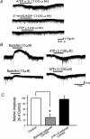
Figure 6. Effect of P2X agonist and antagonists
A, ATPγS (100 μm) induced an inward current (upper trace), while αβmeATP (100 μm), a P2X receptor agonist, did not cause any inward current in the same lamina V neurones (lower trace). B, TNP-ATP (1 μm), a P2X receptor antagonist, did not affect the ATPγS-induced inward current (middle trace). On the other hand, in the presence of PPADS (10 μm), the ATPγS-induced inward current was completely abolished in the same neurons (lower trace). C, a summary of the averaged amplitude of the inward currents by αβmeATP (100 μm), ATPγS (100 μm), ATPγS (100 μm) in the presence of TNP-ATP (1 μm), and ATPγS (100 μm) in the presence of PPADS (10 μm). The ATPγS-induced inward currents in the presence of PPADS were significantly smaller than those in the absence of PPADS (*P < 0.05). D, a summary of the relative sEPSC frequency before (control) and during the application of αβmeATP (100 μm), ATPγS (100 μm), ATPγS (100 μm) in the presence of TNP-ATP (1 μm), and ATPγS (100 μm) in the presence of PPADS (10 μm). Both αβmeATP and ATPγS significantly increased sEPSC frequency (*P < 0.05). The ATPγS-induced increase in sEPSC frequency was not significantly inhibited by TNP-ATP, but was markedly suppressed by PPADS (*P < 0.05). Error bars s.e.m.
Figure 7
Figure 7. Developmental change of postsynaptic P2X receptors
A, ATPγS (100 μm) did not produce any membrane current in a P9–12 lamina V neurone. B, ATPγS (100 μm) induced a clear inward current in a P16–19 lamina V neurone. In contrast to P9–12 lamina V neurones, the ATPγS-induced inward current was observed in all P16–19 lamina V neurones examined. C, a summary of the incidence of the ATPγS-induced inward currents in P9–12 and P16–19 lamina V neurones. The population of P16–19 lamina V neurones which exhibited the inward current was significantly larger than that of P9–12 lamina V neurones (*P < 0.05). D, a summary of the averaged amplitude of the ATPγS-induced inward currents in P9–12 and P16–19 lamina V neurones at these stages. The average amplitude of the ATPγS-induced inward currents in P16–19 was significantly larger than that in P9–12 (*P < 0.05). Error bars s.e.m.

Similar articles

Cited by

References

    1. Anand KJ. Effects of perinatal pain and stress. Prog Brain Res. 2000;122:117–129. - PubMed
    1. Bardoni R, Goldstein PA, Lee CJ, Gu JG, MacDermott AB. ATP P2X receptors mediate fast synaptic transmission in the dorsal horn of the rat spinal cord. J Neurosci. 1997;17:5297–5304. - PMC - PubMed
    1. Burnstock G, Wood JN. Purinergic receptors: their role in nociception and primary afferent neurotransmission. Curr Opin Neurobiol. 1996;6:526–532. - PubMed
    1. Chen M, Gu JG. A P2X receptor-mediated nociceptive afferent pathway to lamina I of the spinal cord. Mol Pain. 2005;1:4. - PMC - PubMed
    1. Chizh BA, Illes P. P2X receptors and nociception. Pharmacol Rev. 2001;53:553–568. - PubMed

Publication types

MeSH terms

Substances

LinkOut - more resources