Comparative Study
. 2006 Jun 15;573(Pt 3):595-609.
doi: 10.1113/jphysiol.2006.107391. Epub 2006 Apr 13.

cAMP sensor Epac as a determinant of ATP-sensitive potassium channel activity in human pancreatic beta cells and rat INS-1 cells

Affiliations
Comparative Study

cAMP sensor Epac as a determinant of ATP-sensitive potassium channel activity in human pancreatic beta cells and rat INS-1 cells

Guoxin Kang et al. J Physiol. .

Abstract

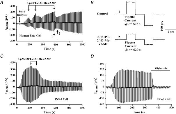
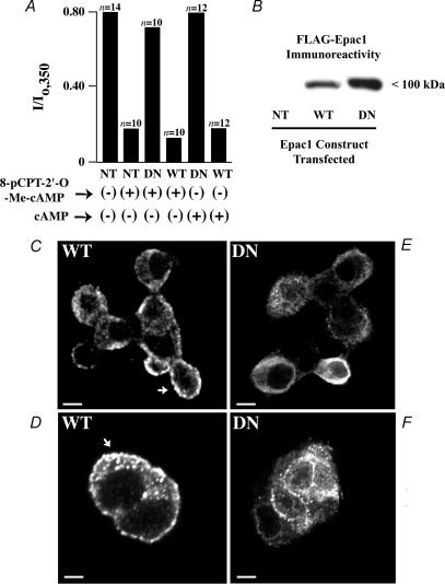
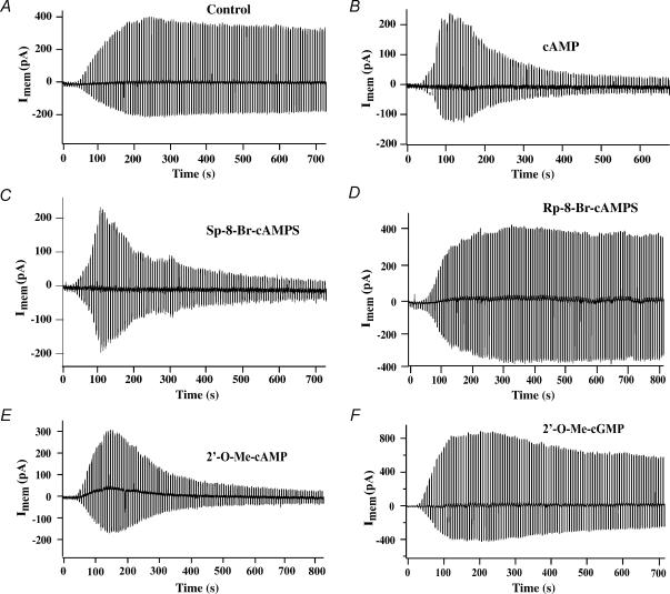
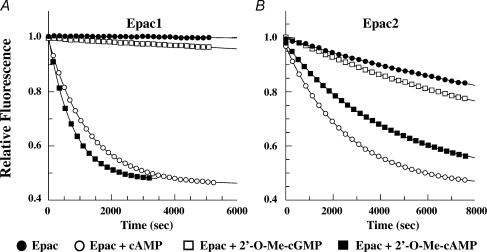
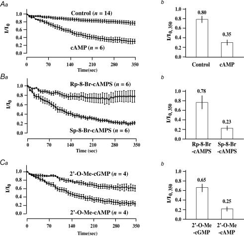
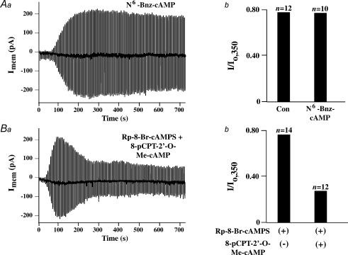
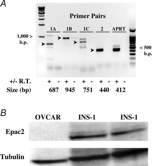
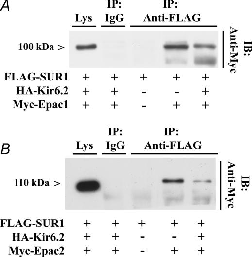
The Epac family of cAMP-regulated guanine nucleotide exchange factors (cAMPGEFs, also known as Epac1 and Epac2) mediate stimulatory actions of the second messenger cAMP on insulin secretion from pancreatic beta cells. Because Epac2 is reported to interact in vitro with the isolated nucleotide-binding fold-1 (NBF-1) of the beta-cell sulphonylurea receptor-1 (SUR1), we hypothesized that cAMP might act via Epac1 and/or Epac2 to inhibit beta-cell ATP-sensitive K+ channels (K(ATP) channels; a hetero-octomer of SUR1 and Kir6.2). If so, Epac-mediated inhibition of K(ATP) channels might explain prior reports that cAMP-elevating agents promote beta-cell depolarization, Ca2+ influx and insulin secretion. Here we report that Epac-selective cAMP analogues (2'-O-Me-cAMP; 8-pCPT-2'-O-Me-cAMP; 8-pMeOPT-2'-O-Me-cAMP), but not a cGMP analogue (2'-O-Me-cGMP), inhibit the function of K(ATP) channels in human beta cells and rat INS-1 insulin-secreting cells. Inhibition of K(ATP) channels is also observed when cAMP, itself, is administered intracellularly, whereas no such effect is observed upon administration N6-Bnz-cAMP, a cAMP analogue that activates protein kinase A (PKA) but not Epac. The inhibitory actions of Epac-selective cAMP analogues at K(ATP) channels are mimicked by a cAMP agonist (8-Bromoadenosine-3', 5'-cyclic monophosphorothioate, Sp-isomer, Sp-8-Br-cAMPS), but not a cAMP antagonist (8-Bromoadenosine-3', 5'-cyclic monophosphorothioate, Rp-isomer, Rp-8-Br-cAMPS), and are abrogated following transfection of INS-1 cells with a dominant-negative Epac1 that fails to bind cAMP. Because both Epac1 and Epac2 coimmunoprecipitate with full-length SUR1 in HEK cell lysates, such findings delineate a novel mechanism of second messenger signal transduction in which cAMP acts via Epac to modulate ion channel function, an effect measurable as the inhibition of K(ATP) channel activity in pancreatic beta cells.


Figures

Figure 1
Figure 1. Inhibition of the whole-cell KATP current by Epac-selective cAMP analogues
A and B, 8-pCPT-2′-O-Me-cAMP (100 μm; individual 30 s applications indicated by vertical arrows) inhibited the KATP current of a human β cell, as depicted on a compressed (A) and an expanded (B) time scale. Time points 1 and 2 in A are illustrated in B as the pipette currents measured at t = 575 and t = 620 s. Outward currents are indicated by upward current deflections. The membrane conductances at t = 575 and t = 620 s, respectively, were 10.0 and 4.7 nS. C, the KATP current of an INS-1 cell was inhibited by 8-pMeOPT-2′-O-Me-cAMP (300 μm; individual 30 s applications indicated by arrows). D, glyburide (10 nm, continual application indicated by the horizontal line) abolished the KATP current of an INS-1 cell. All test solutions were applied extracellularly to individual cells using a ‘puffer’ pipette.
Figure 6
Figure 6. Dominant-negative Epac1 diminishes the action of 8-pCPT-2′-O-Me-cAMP
A, the amplitude of the normalized whole-cell KATP current of INS-1 cells measured at t = 350 s (I/Io,350) is illustrated for INS-1 cells not transfected (NT) or stably transfected with either wild-type (WT) FLAG-Epac1 or dominant-negative (DN) FLAG-Epac1. Cells dialysed with 8-pCPT-2′-O-Me-cAMP (100 μm) or cAMP (300 μm) are indicated as (+), whereas cells not dialysed with 8-pCPT-2′-O-Me-cAMP or cAMP are indicated as (−). B, expression of WT and DN FLAG-tagged Epac1 in lysates of stably transfected INS-1 cells was confirmed by immunoblot analysis, whereas no such immunoreactivity was detected in INS-1 cells not transfected (NT). CF, immunofluorescence cytochemistry for detection of WT and DN FLAG-Epac1 in stably transfected INS-1 cells. Punctate immunoreactivity (arrows) corresponding to WT or DN FLAG-Epac1 was apparent at the plasma membrane and in the cytoplasm of a cluster of cells (C, E and F) or a pair of cells (D). No such immunoreactivity was detected in cells not transfected with FLAG-Epac1. Calibration bars: 3.4 μm for C, E and F; 2.5 μm for D.
Figure 2
Figure 2. Inhibition of the KATP current of INS-1 cells by intracellularly applied cyclic nucleotide analogues
A, dialysis with a pipette solution containing no cyclic nucleotide resulted in the appearance of the whole-cell KATP current under control conditions. B, inclusion of cAMP (300 μm) in the patch pipette did not prevent the appearance of the KATP current once dialysis had commenced at t = 0, but it resulted in a prompt decrease of the KATP current measured at later time points. CF, the action of cAMP was reproduced by 100 μm each of Sp-8-Br-cAMPS (C) and 2′-O-Me-cAMP (E), but not Rp-8-Br-cAMPS (D) or 2′-O-Me-cGMP (F).
Figure 3
Figure 3. cAMP and 2′-O-Me-cAMP but not 2′-O-Me-cGMP act via Epac to stimulate guanyl nucleotide exchange on Rap1
Rap1B loaded with a fluorescent GDP analogue was incubated in the presence of non-fluorescent GDP and either Epac1 (A) or Epac2 (B). cAMP, 2′-O-Me-cAMP or 2′-O-Me-cGMP was then added at a final concentration of 500 μm. Exchange of fluorescent GDP for non-fluorescent GDP was measured spectrophotometrically in real time. When bound to Rap1B, fluorescent GDP exhibits greater fluorescence than when it is simply dissolved in buffer solution. Thus, the decay of fluorescence measured in this assay reflects Epac-mediated stimulation of guanyl nucleotide exchange on Rap1B. Note that when tested at a saturating concentration (500 μm), the Epac1-mediated action of 2′-O-Me-cAMP proceeds at a faster rate than that mediated by Epac2.
Figure 4
Figure 4. Comparison of the rates of KATP current decay measured under conditions in which INS-1 cells were dialysed with a pipette solution containing cyclic nucleotides
Aa, Ba and Ca, the time course of whole-cell KATP current decay is illustrated under control conditions, or conditions in which the pipette solution contained cyclic nucleotides. The current measured at t = 0 is the maximal current (Io) achieved following the initiation of dialysis. The current (I) measured at subsequent time points is normalized relative to Io. Values of n correspond to the number of cells studied under each experimental condition. Ab, Bb and Cb, the amplitude of the normalized KATP current measured at t = 350 s (I/Io,350) is illustrated under control conditions, or conditions in which the pipette solution contained cyclic nucleotides. All cyclic nucleotides were administered at a concentration of 100 μm, except for cAMP (300 μm). Error bars indicate means ±s.e.m.
Figure 5
Figure 5. Assessment of the actions of N6-Bnz-cAMP, Rp-8-Br-cAMPS and 8-pCPT-2′-O-Me-cAMP in INS-1 cells
A, inclusion of N6-Bnz-cAMP (100 μm) in the patch pipette solution was without effect on the rate of decay (Aa) or absolute magnitude (Ab) of the KATP current measured under conditions of whole-cell dialysis. See the legend of Fig. 4 for an explanation of how the value of I/Io,350 was calculated. B, inclusion of Rp-8-Br-cAMPS (100 μm) in the patch pipette solution failed to influence the action of 8-pCPT-2′-O-Me-cAMP (100 μm) to increase the rate of decay of the KATP current (Ba), and to decrease its absolute magnitude (Bb).
Figure 7
Figure 7. Expression of endogenous Epac in INS-1 cells
A, RT-PCR was performed using three sets of Epac1 primer pairs (designated as 1A, 1B and 1C) and a single Epac2 primer pair (designated as 2). PCR product size is indicated in base pairs. Arrowheads indicate PCR products of the expected sizes. Abbreviations: +/− R.T., template generated with or without reverse transcriptase added to the cDNA synthesis reaction; APRT, adenine phosphoribosyltransferase control. A PCR product corresponding to APRT can be derived by PCR of cDNA derived from mRNA but not genomic DNA when using the rat APRT forward (5′-TCCGAATCTGAGTTGCAGC-3′) and reverse primers (5′-CTGCACACATGGTTC-CTCC-3′). B, Epac2 was detected in INS-1 cells (two different platings) by Western blot analysis using an anti-Epac2 monoclonal antiserum. No such immunoreactivity was detected in an ovarian carcinoma cell line (OVCAR). Anti-tubulin antiserum was used to verify loading of the wells with equal amounts of proteins derived from whole-cell lysates.
Figure 8
Figure 8. Epac1 and Epac2 interact with SUR1
A, anti-FLAG antiserum affixed to protein A/G Sepharose beads was used to immunoprecipitate FLAG-SUR1 from lysates of HEK cells transfected with FLAG-SUR1, HA-Kir6.2 and myc-Epac1. The immunoprecipitate (IP) was subjected to immunoblot (IB) analysis using anti-myc antiserum. A 100 kDa immunoreactivity corresponding to myc-Epac1 was detected in lysates of cells transfected with FLAG-SUR1 and myc-Epac1, but not in lysates obtained from cells transfected with FLAG-SUR1 only. Control experiments demonstrated that myc-Epac1 was expressed in whole-cell lysates (Lys), whereas no immunoreactivity was detected in lysates subjected to immunoprecipitation with non-specific rabbit IgG. B, identical findings to those presented in A were obtained when HEK cells were transfected with myc-Epac2 appearing as 115 kDa immunoreactivity. Lower bands correspond to non-specific myc immunoreactivity.

Similar articles

Cited by

References

    1. Aguilar-Bryan L, Nichols CG, Wechsler SW, Clement JP, 4th, Boyd AE, 3rd, Gonzalez G, Herrera-Sosa H, Nguy K, Bryan J, Nelson DA. Cloning of the beta cell high-affinity sulfonylurea receptor: a regulator of insulin secretion. Science. 1995;268:423–426. - PubMed
    1. Asfari M, Janjic D, Meda P, Li G, Halban PA, Wollheim CB. Establishment of 2-mercaptoethanol-dependent differentiated insulin-secreting cell lines. Endocrinology. 1992;130:167–178. - PubMed
    1. Ashcroft FM. ATP-sensitive potassium channelopathies: focus on insulin secretion. J Clin Invest. 2005;115:2047–2058. - PMC - PubMed
    1. Barnett DW, Pressel DM, Chern HT, Scharp DW, Misler S. cAMP-enhancing agents ‘permit’ stimulus–secretion coupling in canine pancreatic islet beta-cells. J Membr Biol. 1994;138:113–120. - PubMed
    1. Baukrowitz T, Schulte U, Oliver D, Herlitze S, Krauter T, Tucker SJ, Ruppersberg JP, Fakler B. PIP2 and PIP as determinants for ATP inhibition of KATP channels. Science. 1998;282:1141–1144. - PubMed

Publication types

MeSH terms

Substances

LinkOut - more resources