

Endothelial cell proliferation in the choriocapillaris during human retinal differentiation
- PMID: 16613918
- PMCID: PMC1857208
- DOI: 10.1136/bjo.2006.092080
Endothelial cell proliferation in the choriocapillaris during human retinal differentiation
Abstract
Background: Differentiation patterns of the neural retina and its retinal vasculature are not well matched. The foveal region differentiates first, however the central retina is not vascularised until late in gestation. The authors explored the hypothesis that higher rates of endothelial cell proliferation in the choriocapillaris of the central retina might compensate for the slow growth of central retinal vessels, providing supplementary nutrients to the region during the early stages of neuronal maturation.
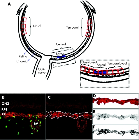
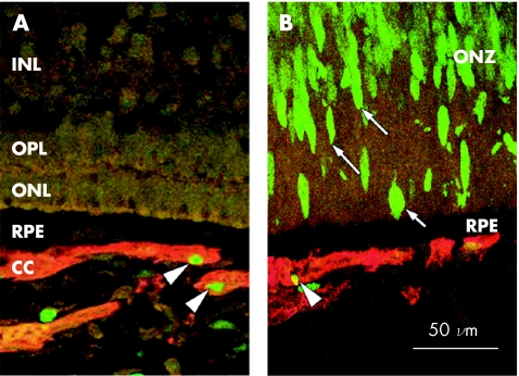
Methods: Frozen sections of five human fetal eyes (14-18.5 weeks' gestation), were examined for Ki-67 and CD34 immunoreactivity using confocal microscopy. Measurements of choriocapillaris area and the number of proliferating choroidal endothelial cells were used to calculate the rate of choroidal endothelial proliferation at five different chorioretinal locations.
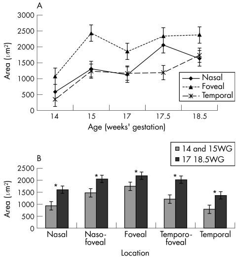
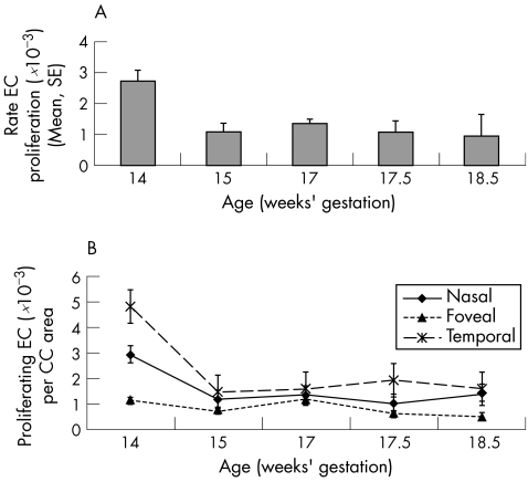
Results: The choriocapillaris area is consistently greater in the foveal region than at other locations and increases progressively with age. A higher rate of endothelial cell proliferation was found in parts of the choriocapillaris associated with the undifferentiated (proliferating) neural retina, compared with the differentiated, central region.
Conclusion: The findings suggest that mechanisms regulating proliferation and growth of the choroidal vasculature are independent of differentiation in the neural retina, and are thus profoundly different from mechanisms that regulate formation of the retinal vasculature.
Similar articles
-
Development of the hyaloid, choroidal and retinal vasculatures in the fetal human eye.Prog Retin Eye Res. 2018 Jan;62:58-76. doi: 10.1016/j.preteyeres.2017.10.001. Epub 2017 Nov 2. Prog Retin Eye Res. 2018. PMID: 29081352 Free PMC article. Review.
-
[A new approach for studying the retinal and choroidal circulation].Nippon Ganka Gakkai Zasshi. 2004 Dec;108(12):836-61; discussion 862. Nippon Ganka Gakkai Zasshi. 2004. PMID: 15656089 Review. Japanese.
-
The initial fetal human retinal vasculature develops by vasculogenesis.Dev Dyn. 2006 Dec;235(12):3336-47. doi: 10.1002/dvdy.20988. Dev Dyn. 2006. PMID: 17061263 Free PMC article.
-
Astrocyte proliferation during development of the human retinal vasculature.Exp Eye Res. 1999 Nov;69(5):511-23. doi: 10.1006/exer.1999.0730. Exp Eye Res. 1999. PMID: 10548471
-
Synchronized tissue-scale vasculogenesis and ubiquitous lateral sprouting underlie the unique architecture of the choriocapillaris.Dev Biol. 2020 Jan 15;457(2):206-214. doi: 10.1016/j.ydbio.2019.02.002. Epub 2019 Feb 21. Dev Biol. 2020. PMID: 30796893
Cited by
-
Complement activation and choriocapillaris loss in early AMD: implications for pathophysiology and therapy.Prog Retin Eye Res. 2015 Mar;45:1-29. doi: 10.1016/j.preteyeres.2014.11.005. Epub 2014 Dec 5. Prog Retin Eye Res. 2015. PMID: 25486088 Free PMC article. Review.
-
Cellular and physiological mechanisms underlying blood flow regulation in the retina and choroid in health and disease.Prog Retin Eye Res. 2012 Sep;31(5):377-406. doi: 10.1016/j.preteyeres.2012.04.004. Epub 2012 May 3. Prog Retin Eye Res. 2012. PMID: 22580107 Free PMC article. Review.
-
Development of the hyaloid, choroidal and retinal vasculatures in the fetal human eye.Prog Retin Eye Res. 2018 Jan;62:58-76. doi: 10.1016/j.preteyeres.2017.10.001. Epub 2017 Nov 2. Prog Retin Eye Res. 2018. PMID: 29081352 Free PMC article. Review.
References
-
- Ozanics V, Rayborn M E, Sagun D. Observations on the ultrastructure of the developing primate choroid coat. Exp Eye Res 19782625–45. - PubMed
-
- Heimann K. The development of the choroid in man. Ophthalmic Res 19723257–273.
-
- Michaelson I C. The mode of development of the vascular system of the retina, with some observations on its significance for certain retinal diseases. Trans Ophthalmol Soc UK 194868137–181.
-
- Nilhausen K. The vasoformative tissue in the foetal retina with particular reference to the histochemical demonstration of its alkaline phosphatase activity. Acta Ophthalmol 19583665–70. - PubMed
Publication types
MeSH terms
LinkOut - more resources
Full Text Sources