. 2006 Apr 25;103(17):6489-94.
doi: 10.1073/pnas.0509531103. Epub 2006 Apr 13.

S-adenosylmethionine stabilizes cystathionine beta-synthase and modulates redox capacity

Affiliations

S-adenosylmethionine stabilizes cystathionine beta-synthase and modulates redox capacity

Anna Prudova et al. Proc Natl Acad Sci U S A. .

Abstract

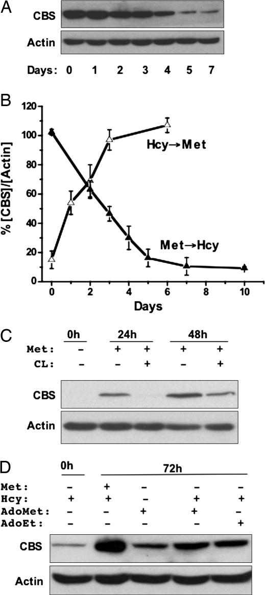
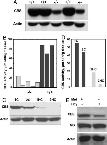
The transsulfuration pathway converts homocysteine to cysteine and represents the metabolic link between antioxidant and methylation metabolism. The first and committing step in this pathway is catalyzed by cystathionine beta-synthase (CBS), which is subject to complex regulation, including allosteric activation by the methyl donor, S-adenosylmethionine (AdoMet). In this study, we demonstrate that methionine restriction leads to a >10-fold decrease in CBS protein levels, and pulse proteolysis studies reveal that binding of AdoMet stabilizes the protein against degradation by approximately 12 kcal/mol. These observations predict that under pathological conditions where AdoMet levels are diminished, CBS, and therefore glutathione levels, will be reduced. Indeed, we demonstrate this to be the case in a mouse model for spontaneous steatohepatitis in which the gene for the MAT1A isoenzyme encoding AdoMet synthetase has been disrupted, and in human hepatocellular carcinoma, where MAT1A is silenced. Furthermore, diminished CBS levels are associated with reduced cell viability in hepatoma cells challenged with tert-butyl hydroperoxide. This study uncovers a mechanism by which CBS is allosterically activated by AdoMet under normal conditions but is destabilized under pathological conditions, for redirecting the metabolic flux toward methionine conservation. A mechanistic basis for the coordinate changes in redox and methylation metabolism that are a hallmark of several complex diseases is explained by these observations.


Conflict of interest statement

Conflict of interest statement: No conflicts declared.

Figures

Fig. 1.
Fig. 1.
Methionine metabolism in mammals. CBS, MS, CGL, PPG, and MAT refer to cystathionine β-synthase, methionine synthase, γ-cystathionase, propargylglycine, and methionine adenosyltransferase, respectively. The inhibitors used to block specific enzymes are indicated. AdoHcy, S-adenosylhomocysteine.
Fig. 2.
Fig. 2.
Regulation of CBS by methionine status. (A) SkHep-1 cells were grown for 7 days in MEM containing 100 μM methionine and switched to MetHcy+ medium containing 200 μM dl-homocysteine. CBS and actin (an equal loading control) were detected in cell extracts by Western analysis as described under Methods and are representative of four independent experiments. A similar response was observed in the human hepatoma cell line HepG2 and the transformed monkey kidney cell line COS-1 (not shown). (B) Reversible regulation of CBS by methionine. Change in CBS concentration normalized to the density of actin in each lane was plotted as a function of time in cells switched from Met+ to MetHcy+ medium or vice versa. Before switching to Met+ medium, SKHep-1 cells were cultured for 7 days in MetHcy+ medium. Data are the average of four independent experiments. (C) AdoMet signals changes in methionine status. SkHep-1 cells were grown in MetHcy+ medium for 7 days, CBS expression was induced by transferring cells to Met+ medium with or without 20 mM cycloleucine (CL), and CBS levels were assessed by Western analysis 0, 24, and 48 h later. Data are representative of at least three experiments performed in duplicate. (D) AdoMet or its structural analog, AdoEt, can induce CBS under conditions of methionine restriction. SkHep-1 cells grown for 7 days in MetHcy+ medium were transferred to the same medium supplemented with either 100 μM methionine or 500 μM AdoMet or 500 μM AdoEt. CBS levels were assessed by Western blot analysis 72 h later. Data are representative of two experiments performed in duplicate.
Fig. 3.
Fig. 3.
CBS expression in liver disease states. (A) Hepatic CBS levels were determined by Western blot analysis in wild type (+/+) and MAT1A knockout (−/−) mice. Protein concentration was determined by using the Bradford assay, and equal loading of samples in each lane was ensured by monitoring actin levels. (B) CBS-specific activity was determined by the ninhydrin assay (8) in wild-type (black bars) and MAT1A knockout (gray bars) mouse livers. (C) Hepatic CBS levels were determined by Western blot analysis in control (1C and 2C) and cancerous (1HC and 2HC) biopsied liver tissue from two patients with hepatocellular carcinoma. (D) CBS-specific activity was determined by the ninhydrin assay in normal (black bars) and cancerous (gray bars) liver tissue. Samples 1C and 1HC and 2C and 2HC are derived from normal and hepatocellular cancer tissue from the same patient, respectively. (E) Effect of methionine availability on CBS expression in fibroblasts (F4875) homozygous for the D444N mutation in CBS. The patient fibroblast cell line was grown for 7 days in either Met+ or MetHcy+ medium. Levels of CBS, methionine synthase, and actin were monitored by Western analysis. Data are representative of three independent experiments performed in duplicate.
Fig. 4.
Fig. 4.
AdoMet stabilizes CBS. (A and B) Proteolytic digestion of CBS in the absence (A) or presence (B) of 300 μM AdoMet by pulse proteolysis as described in Methods. The first lane in each gel represents molecular-weight markers. (C) Dependence of the fraction of folded protein (ffold) on the denaturant concentration in the absence (open squares) and presence (filled squares) of 300 μM AdoMet. Data are averages of four independent experiments.
Fig. 5.
Fig. 5.
The effect of diminished CBS on cell viability at increasing concentrations of tert-butyl hydroperoxide in cells grown in the Met+ (filled circles), MetHcy+ (filled triangles), or MetHcy+ plus N-acetylcysteine (NAC) (open triangles) medium. Viability is represented as a percentage of viable cells in untreated controls, and each data point is the average of three to six independent determinations.

References

    1. Jungst C., Cheng B., Gehrke R., Schmitz V., Nischalke H. D., Ramakers J., Schramel P., Schirmacher P., Sauerbruch T., Caselmann W. H. Hepatology. 2004;39:1663–1672. - PubMed
    1. McKillop I. H., Schrum L. W. Alcohol. 2005;35:195–203. - PubMed
    1. Cai J., Mao Z., Hwang J. J., Lu S. C. Cancer Res. 1998;58:1444–1450. - PubMed
    1. Finkelstein J. D., Kyle W. E., Martin J. J., Pick A.-M. Biochem. Biophys. Res. Commun. 1975;66:81–87. - PubMed
    1. Taoka S., Widjaja L., Banerjee R. Biochemistry. 1999;38:13155–13161. - PubMed

Publication types

MeSH terms

LinkOut - more resources