Review
. 2006 Apr;19(2):338-81.
doi: 10.1128/CMR.19.2.338-381.2006.

The continuing challenges of leprosy

Affiliations
Review

The continuing challenges of leprosy

D M Scollard et al. Clin Microbiol Rev. 2006 Apr.

Abstract

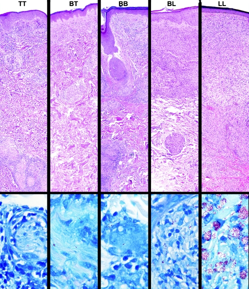
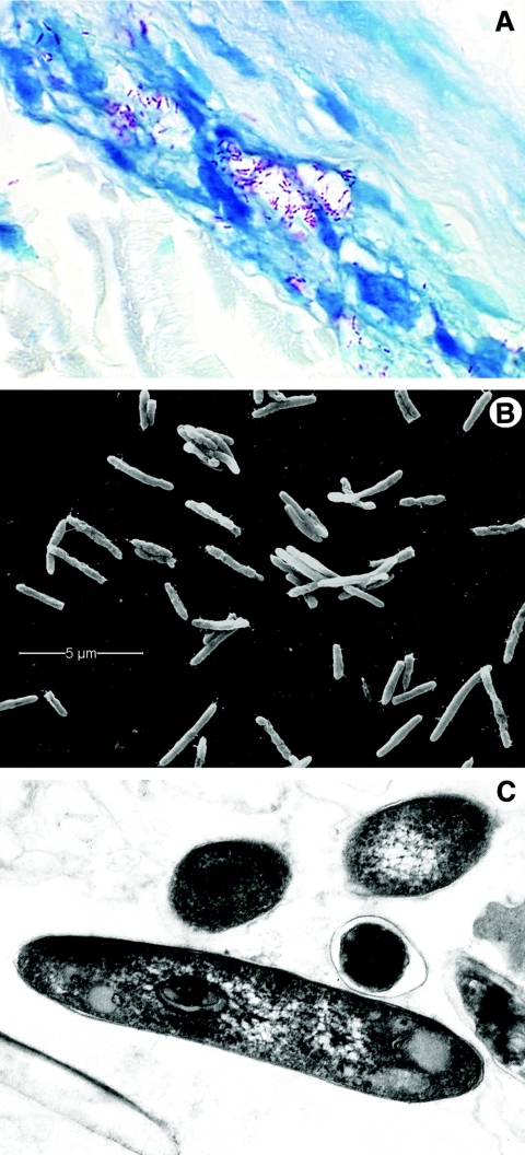
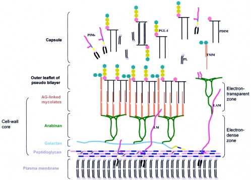
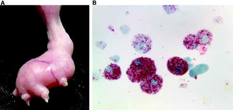
Leprosy is best understood as two conjoined diseases. The first is a chronic mycobacterial infection that elicits an extraordinary range of cellular immune responses in humans. The second is a peripheral neuropathy that is initiated by the infection and the accompanying immunological events. The infection is curable but not preventable, and leprosy remains a major global health problem, especially in the developing world, publicity to the contrary notwithstanding. Mycobacterium leprae remains noncultivable, and for over a century leprosy has presented major challenges in the fields of microbiology, pathology, immunology, and genetics; it continues to do so today. This review focuses on recent advances in our understanding of M. leprae and the host response to it, especially concerning molecular identification of M. leprae, knowledge of its genome, transcriptome, and proteome, its mechanisms of microbial resistance, and recognition of strains by variable-number tandem repeat analysis. Advances in experimental models include studies in gene knockout mice and the development of molecular techniques to explore the armadillo model. In clinical studies, notable progress has been made concerning the immunology and immunopathology of leprosy, the genetics of human resistance, mechanisms of nerve injury, and chemotherapy. In nearly all of these areas, however, leprosy remains poorly understood compared to other major bacterial diseases.


Figures

FIG. 1.
FIG. 1.
Immunopathologic spectrum of leprosy. Representative fields from each of the histopathological types of leprosy in the Ridley-Jopling classification are presented in the upper panel, in hematoxylin- and eosin-stained sections (magnification, ×63). The well-formed epithelioid granulomatous infiltrates seen in polar tuberculoid (TT) lesions become increasingly disorganized in each successive increment in the scale until they become completely disorganized aggregates of foamy histiocytes, with only occasional lymphocytes, in polar lepromatous (LL) lesions. Representative fields of each classification are shown in Fite-stained sections in the lower panel (magnification, ×1,000). A search of more than 50 fields was required to find the two organisms shown in a cutaneous nerve in the TT sample, and organisms are often similarly difficult to find in BT lesions. This spectrum is the yardstick against which is measured each new hypothesis and discovery regarding immunological mechanisms proposed to be responsible for the wide range of human responses to M. leprae.
FIG. 2.
FIG. 2.
Morphology of M. leprae. A. M. leprae is weakly acid fast but, when stained with the Fite-Faraco method, it appears as red, rod-shaped organisms; shorter beaded or granular shapes are observed when the bacilli are dead or dying. The organisms are seen here within a human nerve, counterstained with methylene blue. Magnification, approximately ×800. B. A suspension of nude-mouse footpad-derived M. leprae under the scanning electron microscope, which reveals the surface of the organisms. M. leprae, like other mycobacteria, tends to cluster. Magnification, approximately ×12,000. C. Internal features of M. leprae are observed in this ultrathin section of the bacilli under a transmission electron microscope. The round and oval images seen in the upper portion of this photograph are bacilli that have been cut in cross section. Magnification, ×29,000.
FIG. 3.
FIG. 3.
Schematic model of the cell envelope of M. leprae. The plasma membrane is covered by a cell wall core made of peptidoglycan covalently linked to the galactan by a linker unit of arabinogalactan. Three branched chains of arabinan are in turn linked to the galactan. Mycolic acids are linked to the termini of the arabinan chains to form the inner leaflet of a pseudolipid bilayer. An outer leaflet is formed by the mycolic acids of trehalose monomycolates (TMM) and mycocerosoic acids of phthiocerol dimycocerosates (PDIMs) and PGLs as shown. A capsule presumably composed largely of PGLs and other molecules such as PDIMs, phosphatidylinositol mannosides, and phospholipids surrounds the bacterium. Lipoglycans such as phosphatidylinositol mannosides, lipomannan (LM), and lipoarabinomannan (LAM), known to be anchored in the plasma membrane, are also found in the capsular layer as shown. (Reprinted from reference with permission of the publisher.)
FIG. 4.
FIG. 4.
Cultivationof M. leprae in mouse footpads. A. Enlarged nude-mouse footpad 6 months after infection with 5 × 10⁷ live M. leprae. B. Heavily infected macrophages harvested from mouse footpad (magnification, ×1,000).
FIG. 5.
FIG. 5.
Two-stage model of genetic influence on human immunity to M. leprae. Infection by M. leprae probably occurs through a skin or nasal route, by mechanisms that are not yet defined. The various genes and loci listed are discussed in the text under Genetic Influences, and the cells listed are discussed in the succeeding sections.
FIG. 6.
FIG. 6.
Proposed model of infection of peripheral nerve by M. leprae via blood vessels. A cutaneous nerve with three fascicles is represented here to illustrate the proposed steps in the pathogenesis of infection of peripheral nerves by M. leprae. (A) Initially, colonization of the epineurium (e) occurs when bacilli (red) localize in cells in and around blood vessels (blue). It is possible that this is enhanced by drainage of bacilli through the lymphatics (green) that are intertwined with the blood vessels of the epineurium (lymphatics are here illustrated only at the lower end of the drawing). The resulting accumulation of bacilli within and around endothelial cells greatly increases the likelihood that bacilli will be available for circulation through the endoneurial vessels which branch off the epineurial ones. (B) Entry of M. leprae into the endoneurial compartment proceeds along blood vessels from foci on and within the perineurium (p), extending through it into the interior of the nerve. The mechanisms responsible for entry into the interstitial space of the endoneurium remain to be determined. Once inside, however, bacilli are available for phagocytosis by Schwann cells (SC), represented here with concentric layers of myelin surrounding axons. Although these initial events in the localization and entry of M. leprae into peripheral nerves are postulated to be unrelated to specific immune function, the subsequent pathogenesis of neuritis in leprosy probably depends in large part on the patient's immune response to M. leprae. (C) If no effective immune respose develops (e.g., lepromatous leprosy), bacilli proliferate within macrophages and Schwann cells. This results in perineurial inflammation and thickening (proliferation) and an increasing bacterial load both in the epineurium and in the endoneurium. Since M. leprae is an indolent, well-adapted intracellular pathogen, however, axons are not badly damaged for a long time, and a variable degree of nerve function is preserved until late in the course of the disease. (D) If effective cellular immunity and delayed hypersensitivity do develop (e.g., tuberculoid leprosy), a granulomatous response follows at sites of infection near epineurial and endoneurial vessels and Schwann cells. This immunologically elicited inflammation eliminates nearly all of the bacilli in the epi- and perineurium and also stimulates perineurial fibrosis and thickening. However, M. leprae organisms that have already been ingested by Schwann cells may be relatively protected from immunologically mediated destruction and able to maintain a persistent infection in these cells for a longer time. This is where most bacilli are found in diagnostic biopsies of tuberculoid lesions. Granulomatous inflammation is also potentially injurious to adjacent tissue. In M. leprae-infected nerves, this includes injury to axons in the vicinity of the granulomas, resulting in impaired nerve function. (Reprinted from reference .)
FIG. 7.
FIG. 7.
Transmission electron micrographs of Mycobacterium leprae-infected rat Schwann cell (SC)-neuron cocultures. Infected cultures were obtained by exposure of primary Schwann cells to M. leprae for 48 h. After cultivation for 12 days at 33°C, they were seeded onto embryonic rat neurons. The infected Schwann cell cultures were induced to myelinate and cultured for 30 days at 33°C. A. Myelinating Schwann cells. B. Nonmyelinating Schwann cells. (Reprinted from reference with permission. © 2002 by the Infectious Diseases Society of America. All rights reserved.)

References

    1. Abel, L., and F. Demenais. 1988. Detection of major genes for susceptibility to leprosy and its subtypes in a Caribbean island: Desirade Island. Am. J. Hum. Genet. 42:256-266. - PMC - PubMed
    1. Abel, L., F. O. Sanchez, J. Oberti, N. V. Thuc, L. V. Hoa, V. D. Lap, E. Skamene, P. H. Lagrange, and E. Schurr. 1998. Susceptibility to leprosy is linked to the human NRAMP1 gene. J. Infect. Dis. 177:133-145. - PubMed
    1. Adams, L. B., M. C. Dinauer, D. E. Morgenstern, and J. L. Krahenbuhl. 1997. Comparison of the roles of reactive oxygen and nitrogen intermediates in the host response to Mycobacterium tuberculosis using transgenic mice. Tubercle Lung Dis. 7 8:237-246. - PubMed
    1. Adams, L. B., S. G. Franzblau, Z. Vavrin, J. B. Hibbs, Jr., and J. L. Krahenbuhl. 1991. l-Arginine-dependent macrophage effector functions inhibit metabolic activity of Mycobacterium leprae. J. Immunol. 147:1642-1646. - PubMed
    1. Adams, L. B., Y. Fukutomi, and J. L. Krahenbuhl. 1993. Regulation of murine macrophage effector functions by lipoarabinomannan from mycobacterial strains with different degrees of virulence. Infect. Immun. 61:4173-4181. - PMC - PubMed

Publication types

MeSH terms