. 2006 Apr 13;34(7):e53.
doi: 10.1093/nar/gkl143.

Polycistronic RNA polymerase II expression vectors for RNA interference based on BIC/miR-155

Affiliations

Polycistronic RNA polymerase II expression vectors for RNA interference based on BIC/miR-155

Kwan-Ho Chung et al. Nucleic Acids Res. .

Abstract

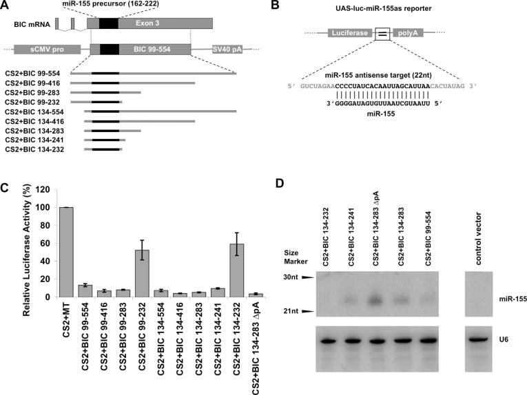
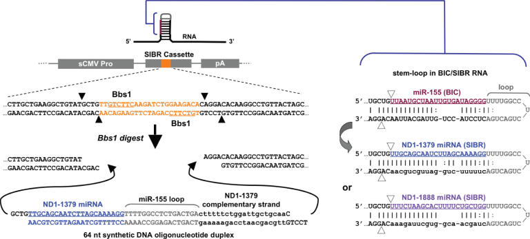
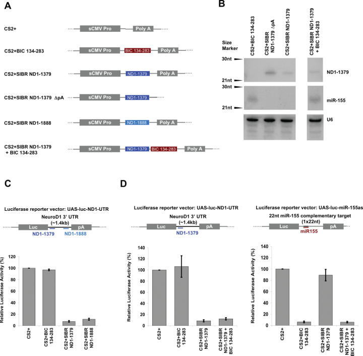
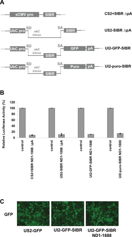
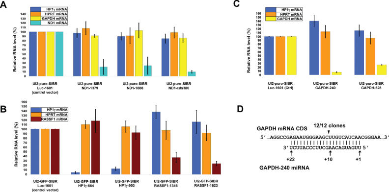
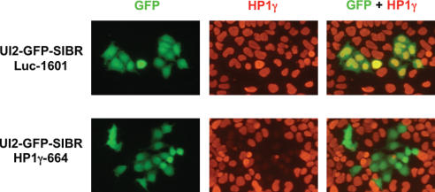
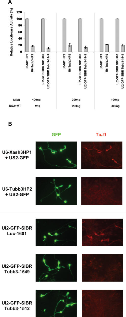
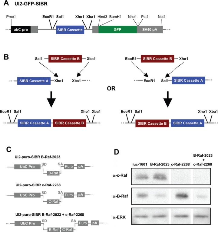
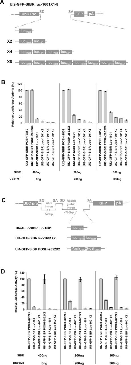
Vector-based RNA interference (RNAi) has emerged as a valuable tool for analysis of gene function. We have developed new RNA polymerase II expression vectors for RNAi, designated SIBR vectors, based upon the non-coding RNA BIC. BIC contains the miR-155 microRNA (miRNA) precursor, and we find that expression of a short region of the third exon of mouse BIC is sufficient to produce miR-155 in mammalian cells. The SIBR vectors use a modified miR-155 precursor stem-loop and flanking BIC sequences to express synthetic miRNAs complementary to target RNAs. Like RNA polymerase III driven short hairpin RNA vectors, the SIBR vectors efficiently reduce target mRNA and protein expression. The synthetic miRNAs can be expressed from an intron, allowing coexpression of a marker or other protein with the miRNAs. In addition, intronic expression of a synthetic miRNA from a two intron vector enhances RNAi. A SIBR vector can express two different miRNAs from a single transcript for effective inhibition of two different target mRNAs. Furthermore, at least eight tandem copies of a synthetic miRNA can be expressed in a polycistronic transcript to increase the inhibition of a target RNA. The SIBR vectors are flexible tools for a variety of RNAi applications.


Figures

Figure 1
Figure 1
Identification of the minimal region of BIC for miR-155 expression. (A) Schematic representation of the mouse BIC non-coding RNA, which contains the miR-155 precursor in the third exon. To express miR-155, 455 nt of the BIC third exon was placed under the control of the sCMV RNA polymerase II promoter, followed by the SV40 late polyadenylation site. Smaller fragments of the third exon (with boundaries based on homologous sequences among human, mouse, and chick BIC RNAs), were used to determine the minimal domain within the third exon of BIC required for miR-155 expression. Positions of starting and ending nucleotides are indicated. Exon and intron sizes are not to scale. (B) The UAS-luc-miR-155as reporter contains a single copy of a 22 nt artificial miR-155 target sequence perfectly complementary to miR-155, inserted into the 3′-UTR of the luciferase mRNA. (C) Relative luciferase activity after cotransfection of BIC expression vectors with the UAS-luc-miR-155as reporter. CS2+MT, which expresses a multimerized myc-epitope tag, was used as a control. BIC 134–283 ΔpA indicates the BIC 134–283 expression vector with the polyadenylation signal deleted. Standard errors are indicated. (D) Upper panel: northern blot for miR-155 in cells transfected with the indicated vectors. The control vector is ND1-1379, in which the miR-155 sequence is replaced with an unrelated sequence (see Figure 2). Lower panel: the same blot re-probed for the U6 snRNA as a loading control.
Figure 2
Figure 2
RNAi vectors based on the miR-155 primary transcript BIC. The SIBR cassette was constructed by replacing the miR-155 precursor stem–loop in the BIC 134–283 fragment with a polylinker (orange) containing inverted Bbs1 restriction sites. The Bbs1 cut sites (filled triangles) are outside the recognition sites (underlined). A synthetic 64 nt DNA duplex can be inserted in which the miR-155 miRNA sequence is replaced by a synthetic miRNA sequence (blue). The primary RNA transcript from the SIBR vector is indicated above the schematic, with the miRNA containing stem–loop indicated and shown in more detail on the right. The stem–loop in the primary RNA is processed by Drosha to release a stem–loop pre-miRNA, which is then processed by Dicer to release the mature miRNA. Expected Drosha cleavage sites are indicated by open triangles. Each mature miRNA is underlined. The miR-155 sequence in the stem–loop of the BIC RNA (red) is replaced by either of two synthetic miRNA sequences (blue or purple) perfectly complementary to the 3′-UTR of the mouse NeuroD1 mRNA (ND1). The complementary strand of the stem–loops for the ND1 miRNAs was altered to maintain a partial duplex with two single base gaps (lower case). The loop (grey) and flanking sequences (upper case black) are from BIC. Numbers for synthetic miRNAs indicate the first nucleotide of the target mRNA that is complementary to the miRNA.
Figure 3
Figure 3
Sequence specific inhibition by SIBR vectors. (A) Schematic diagrams of variants of SIBR expression vectors with single or tandem cassettes. (B) miR-155 and the ND1-1379 synthetic miRNA are detected as single bands and at similar levels by northern blot analysis of RNA from P19 cells transfected with single and tandem SIBR vectors. Deletion of the polyadenylation signal (ND1-1379 ΔpA) increased the level of ND1-1379 miRNA. Bottom panel: the same blot re-probed for the U6 snRNA as a loading control. (C) Cotransfection of SIBR vectors expressing miRNAs complementary to NeuroD inhibit a cotransfected luciferase reporter containing the mouse NeuroD1 3′-UTR, while BIC 134–283 has no effect on luciferase activity. (D) A vector with both a ND1-1379 SIBR cassette and BIC 134–283 in tandem effectively inhibits a luciferase reporter for either miRNA. The level of inhibition from the tandem vector is comparable with that of the individual vectors. Standard errors are indicated.
Figure 4
Figure 4
Expression of the SIBR cassette from an intron. (A) Schematic diagrams of SIBR vectors: US2-SIBR ΔpA vector contains the SIBR cassette in the second exon of human ubC gene, under control of the human ubC promoter, but without a polyA signal. There is no coding region present in either the CS2+SIBR ΔpA or US2-SIBR ΔpA vector. UI2 vectors also use the human ubC promoter but contain the SIBR cassette in the first intron of the ubC gene, with the GFP or puromycin-resistance proteins expressed from the second exon. SD and SA indicate splice donor and splice acceptor, respectively. Exon and intron sizes not to scale. (B) Different SIBR vector designs expressing the ND1-1888 miRNA against NeuroD1 were cotransfected with the NeuroD1 3′-UTR luciferase reporter (see Figure 2). All four designs with ND1-1888 showed similar levels of inhibition of the reporter. Control SIBR vectors used the same designs but expressed an unrelated miRNA directed against the mouse POSH mRNA. Standard errors are indicated. (C) Comparable GFP fluorescence was detected 24 h after transfection with GFP expressed from ubC-based expression vectors, whether or not a functional SIBR cassette was present in the ubC intron.
Figure 5
Figure 5
Knock-down of endogenous genes using UI2 SIBR vectors. (A) qRT–PCR measurements of mRNA levels in P19 cells transiently cotransfected with an expression vector for Ngn2 (to activate endogenous NeuroD1 expression), and various UI2-puro-SIBR vectors. The level of NeuroD1 mRNA was reduced by miRNAs directed against the NeuroD1 3′-UTR (see Figure 2) or coding region (ND1-380), but not by a control miRNA directed against Luciferase. GAPDH and HPRT mRNA levels were not reduced. (B) UI2-GFP-SIBR vectors expressing miRNAs that target the HP1γ or RASSF1 mRNAs reduced the levels of the corresponding endogenous mRNA in transfected P19 cells, but not the HPRT mRNA. (C) UI2-puro-SIBR vectors expressing miRNAs directed against GAPDH reduce endogenous GAPDH mRNA levels. (D) Expression of the GAPDH-240 miRNA leads to cleavage of the endogenous GAPDH mRNA at the expected site (see text). In A–D, transfected cells were selected with puromycin to remove untransfected cells. UI2-GFP-SIBR vectors in B were cotransfected with US2-puro to permit selection.
Figure 6
Figure 6
Reduction of endogenous HP1γ protein in single cells identified by coexpressed GFP. P19 cells transfected with the UI2-GFP-SIBR HP1γ-664 vector express GFP (green) and show reduced HP1γ by indirect immunfluorescence (red) when compared with untransfected cells (no GFP). The HP1γ signal in cells transfected with the UI2-GFP-luc1601 control vector remains unchanged.
Figure 7
Figure 7
Comparable inhibition by SIBR vectors and a U6 shRNA vector. (A) A reporter assay comparing inhibition of a luciferase reporter containing 3′-UTR of mouse tubulin β3 (luc-Tubb3-UTR) cotransfected with either the UI2-GFP-SIBR Tubb3-1549 vector or the U6-Tubb3HP2 vector. At three different plasmid concentrations, both vectors showed comparable levels of inhibition. Standard errors are indicated. (B) Reduction of endogenous mouse tubulin β3 protein at a single cell level demonstrated by immunocytochemistry. U6 or UI2-GFP-SIBR vectors were cotransfected into P19 cells together with the US2-Ngn2 vector to induce neuronal differentiation and β3 tubulin expression. U6 transfections included the US2-GFP vector to label transfected cells. P19 cells transfected with a U6 shRNA vector or either of two UI2-GFP-SIBR vectors (green) directed against β3 tubulin showed substantially reduced tubulin β3 protein (red) by indirect immunfluorescence, relative to cells transfected with control vectors.
Figure 8
Figure 8
Knock-down of two endogenous genes using a single UI2 SIBR vector. (A) Schematic of the UI2-GFP/puro-SIBR vectors showing unique restriction sites flanking the SIBR cassette. Indicated in bold are restriction enzyme sites used for multiplexing SIBR cassettes. (B) Using appropriate restriction enzymes, it is possible to create vectors with tandem SIBR cassettes rapidly. (C) Schematic of vectors with miRNAs directed against the B-Raf and/or c-Raf kinases, including a vector with tandem B-Raf and c-Raf SIBR cassettes. Schematics in A-C are not to scale. (D) Western blot showing reduced levels of either B-Raf or c-Raf protein in cells transfected with SIBR vectors expressing a miRNA against the corresponding mRNA, but not in cells transfected with a control vector expressing a miRNA directed against luciferase. The vector expressing two miRNAs targeting the B-Raf and c-Raf mRNAs reduced the levels of both Raf proteins, but not the ERK kinase.
Figure 9
Figure 9
Increased inhibition by multiple SIBR cassettes expressed from a single intron vector, and by use of a two intron vector. (A) Schematic representation of UI2-GFP-SIBR vectors expressing one to eight tandem copies of the same synthetic miRNA against luciferase. (B) P19 Cells transfected with a fixed amount of target luciferase reporter and a fixed total amount of DNA show dose dependent inhibition of luciferase. At three different DNA concentrations, an increased number of copies of the luc-1601 SIBR cassette in the UI2-GFP vector provided better inhibition. The UI2-GFP-SIBR POSH-2852 control vector expresses a functional synthetic miRNA directed against the mouse POSH gene. A vector with eight copies of the POSH miRNA does not inhibit luciferase. Total DNA amount was kept constant by replacing the SIBR vector with the US2-MT vector, which does not express a miRNA. Standard errors are indicated. (C) Schematics of the two intron UI4-GFP-SIBR vectors. UI4-GFP vectors contain the SIBR cassettes in rabbit globin intron, inserted between the exon 2 and GFP in exon 3. Exons 1 and 2 are noncoding. SD and SA indicate splice donor and splice acceptor, respectively. Approximate intron size is indicated. (D) Cotransfection reporter assay comparing the inhibition by UI4-GFP-SIBR vectors to UI2-GFP-SIBR vectors expressing one or two miRNAs against luciferase. At three different plasmid concentrations, the UI4-GFP-SIBR vectors showed increased inhibition of luciferase relative to the UI2-GFP-SIBR vectors. Standard errors are indicated.

References

    1. Filipowicz W. RNAi: the nuts and bolts of the RISC machine. Cell. 2005;122:17–20. - PubMed
    1. Sandy P., Ventura A., Jacks T. Mammalian RNAi: a practical guide. Biotechniques. 2005;39:215–224. - PubMed
    1. Murchison E.P., Hannon G.J. miRNAs on the move: miRNA biogenesis and the RNAi machinery. Curr. Opin. Cell Biol. 2004;16:223–229. - PubMed
    1. Lagos-Quintana M., Rauhut R., Lendeckel W., Tuschl T. identification of novel genes coding for small expressed RNAs. Science. 2001;294:853–858. - PubMed
    1. Lau N.C., Lim le E.P., Weinstein E.G., Bartel da V.P. An abundant class of tiny RNAs with probable regulatory roles in Caenorhabditis elegans. Science. 2001;294:858–862. - PubMed

Publication types