

A case of autoimmune hemolytic anemia associated with an ovarian teratoma
- PMID: 16614532
- PMCID: PMC2734022
- DOI: 10.3346/jkms.2006.21.2.365
A case of autoimmune hemolytic anemia associated with an ovarian teratoma
Abstract
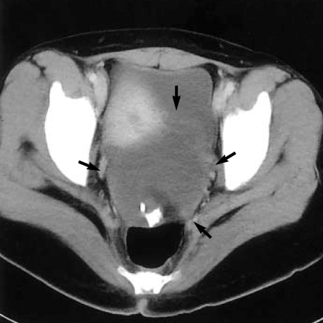
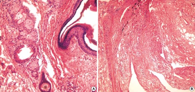
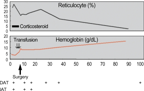
Autoimmune hemolytic anemia associated with an ovarian teratoma is a very rare disease. However, treating teratoma is the only method to cure the hemolytic anemia, so it is necessary to include ovarian teratoma in the differential diagnosis of autoimmune hemolytic anemia. We report herein on a case of a young adult patient who had severe autoimmune hemolytic anemia that was induced by an ovarian teratoma. A 25-yr-old woman complained of general weakness and dizziness for 1 week. The hemoglobin level was 4.2 g/dL, and the direct and indirect antiglobulin tests were all positive. The abdominal computed tomography scan revealed a huge left ovarian mass, and this indicated a teratoma. She was refractory to corticosteroid therapy; however, after surgical resection of the ovarian mass, the hemoglobin level and the reticulocyte count were gradually normalized. The mass was well encapsulated and contained hair and teeth. She was diagnosed as having autoimmune hemolytic anemia associated with an ovarian teratoma. To the best of our knowledge, this is the first such a case to be reported in Korea.
Figures
References
-
- Sallah S, Sigounas G, Vos P, Wan JY, Nguyen NP. Autoimmune hemolytic anemia in patients with non-Hodgkin's lymphoma: characteristics and significance. Ann Oncol. 2000;11:1571–1577. - PubMed
-
- Glorieux I, Chabbert V, Rubie H, Baunin C, Gaspard MH, Guitard J, Duga I, Suc A, Puget C, Robert A. Autoimmune hemolytic anemia associated with a mature ovarian teratoma. Arch Pediatr. 1998;5:41–44. - PubMed
-
- Cobo F, Pereira A, Nomdedeu B, Gallart T, Ordi J, Torne A, Monserrat E, Rozman C. Ovarian dermoid cyst-associated autoimmune hemolytic anemia: a case report with emphasis on pathogenic mechanisms. Am J Clin Pathol. 1996;105:567–571. - PubMed
-
- Buonanno G, Gonnella F, Pettinato G, Castaldo C. Autoimmune hemolytic anemia and dermoid cyst of the mesentery. A case report. Cancer. 1984;54:2533–2536. - PubMed
Publication types
MeSH terms
Substances
LinkOut - more resources
Full Text Sources
Medical
