. 1999 Jan 28;103(4):737-45.
doi: 10.1021/jp9833193.

The effective temperature of Peptide ions dissociated by sustained off-resonance irradiation collisional activation in fourier transform mass spectrometry

Affiliations

The effective temperature of Peptide ions dissociated by sustained off-resonance irradiation collisional activation in fourier transform mass spectrometry

P D Schnier et al. J Phys Chem B. .

Abstract

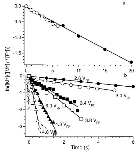
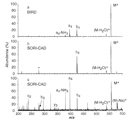
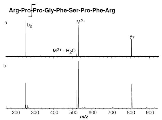
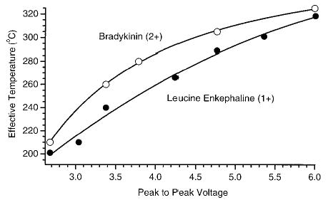
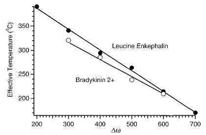
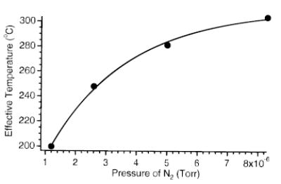
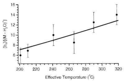
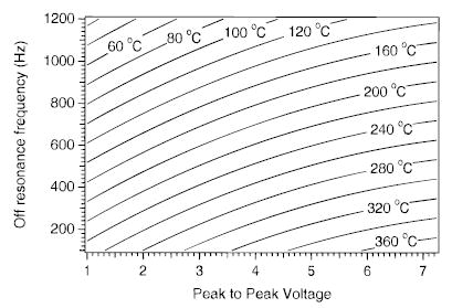
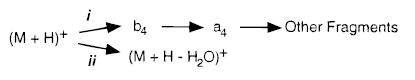
A method for determining the internal energy of biomolecule ions activated by collisions is demonstrated. The dissociation kinetics of protonated leucine enkephalin and doubly protonated bradykinin were measured using sustained off-resonance irradiation (SORI) collisionally activated dissociation (CAD) in a Fourier transform mass spectrometer. Dissociation rate constants are obtained from these kinetic data. In combination with Arrhenius parameters measured with blackbody infrared radiative dissociation, the "effective" temperatures of these ions are obtained. Effects of excitation voltage and frequency and the ion cell pressure were investigated. With typical SORI-CAD experimental conditions, the effective temperatures of these peptide ions range between 200 and 400 degrees C. Higher temperatures can be easily obtained for ions that require more internal energy to dissociate. The effective temperatures of both protonated leucine enkephalin and doubly protonated bradykinin measured with the same experimental conditions are similar. Effective temperatures for protonated leucine enkephalin can also be obtained from the branching ratio of the b(4) and (M + H - H(2)O)(+) pathways. Values obtained from this method are in good agreement with those obtained from the overall dissociation rate constants. Protonated leucine enkephalin is an excellent "thermometer" ion and should be well suited to establishing effective temperatures of ions activated by other dissociation techniques, such as infrared photodissociation, as well as ionization methods, such as matrix assisted laser desorption/ionization.


Figures

Figure 1
Figure 1
Experimental sequence for SORI–CAD experiments.
Figure 2
Figure 2
(a) SORI–CAD spectrum of protonated leucine enkephalin with the excitation frequency applied for 10 ms, 500 Hz below the ions cyclotron frequency (2.5 Vpp). Spectra collected under the same conditions as (a) but with an addition excitation (10Vpp) to continuously remove (b) the b4 ion, (c) (M + H − H2O)+, (d) the a4 ion, and (e) m/z 450 (b–e are double resonance experiments). * indicates a harmonic of the precursor ion.
Figure 3
Figure 3
SORI–CAD dissociation data (ΔΩ = 200 Hz, Vpp = 850 mV) as a function of the time that the nitrogen gas pulse is extended after the irradiation of the ions. The three irradiation times leave the ions at their maximum radius (5.0 mm, 2.5 ms), an intermediate radius (4.4 mm, 3.0 ms), and minimum (unexcited) (5.0 ms) radius.
Figure 4
Figure 4
Normalized precursor ion abundance of protonated leucine enkephalin as a function of SORI excitation time. The open squares and open circles represent data points sampled at times in which the excitation wave form is stopped exactly when the irradiated ions have a maximum and minimum cyclotron radius, respectively.
Figure 5
Figure 5
Dissociation data for protonated leucine enkephalin fit to unimolecular kinetics. (a) Blackbody infrared radiative dissociation kinetics obtained with a cell temperature of 203 °C (○) and SORI–CAD kinetics with an excitation 1200 Hz below the ions cyclotron frequency (2.6 Vpp) (•); (b) SORI–CAD kinetics collected at different excitation voltages (ΔΩ = 1200 Hz, cell pressure = 2.6 × 10⁻⁶ Torr).
Figure 6
Figure 6
Dissociation spectra of protonated leucine enkephalin with (a) BIRD (5 s reaction, 203 °C), and (b) SORI–CAD (ΔΩ = 1200 Hz) with a 2.6Vpp wave form applied for 5 s, and (c) 6.0 Vpp wave form, applied for 10 ms. * indicates a harmonic of the precursor ion.
Figure 7
Figure 7
Spectra of doubly protonated bradykinin dissociated by (a) BIRD (30 s reaction delay, 150 °C), and (b) SORI–CAD (0.5 s reaction delay, ΔΩ = 1200 Hz).
Figure 8
Figure 8
The effective temperature of singly protonated leucine enkephalin and doubly protonated bradykinin ions as a function of the excitation amplitude with ΔΩ = 1200 Hz and a cell pressure of 3 × 10⁻⁶ Torr.
Figure 9
Figure 9
Effective temperature of singly protonated leucine enkephalin and doubly protonated bradykinin ions as a function of the excitation frequency with Vpp = 3 V and a cell pressure of 3 × 10⁻⁶ Torr.
Figure 10
Figure 10
Effective temperature of protonated leucine enkephalin as a function of collision gas pressure during the SORI excite (ΔΩ = 1200 Hz, Vpp = 1.1 V).
Figure 11
Figure 11
Branching ratio of process i:ii (see text) for each SORI–CAD kinetic data set in Figure 5 as a function of Teff. The solid line is the calculated branching ratio from the individual Arrhenius parameters for each process.
Figure 12
Figure 12
Calculated contour plot of the effective temperature of protonated leucine enkephalin as a function of excitation frequency and voltage at a collision gas pressure of 2.6 × 10⁻⁶ Torr. The data were obtained by combining and extrapolating the data shown in Figures 8 and 9.
SCHEME 1
SCHEME 1

References

    1. McLafferty FW. Acc Chem Res. 1994;27:379–386.
    1. Valaskovic GA, Kelleher NL, McLafferty FW. Science. 1996;273:1199–1202. - PubMed
    1. Kelleher NL, Senko MW, Siegel MM, McLafferty FW. J Am Soc Mass Spectrom. 1997;8:380–383.
    1. Williams ER. Anal Chem. 1998;70:179–185A. - PMC - PubMed
    1. Speir JP, Senko MW, Little DP, Loo JA, McLafferty FW. J Mass Spectrom. 1995;30:39–42.

LinkOut - more resources