. 2006 May;116(5):1391-9.
doi: 10.1172/JCI27540. Epub 2006 Apr 13.

Cyclooxygenases, microsomal prostaglandin E synthase-1, and cardiovascular function

Affiliations

Cyclooxygenases, microsomal prostaglandin E synthase-1, and cardiovascular function

Yan Cheng et al. J Clin Invest. 2006 May.

Abstract

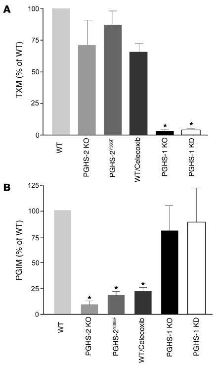
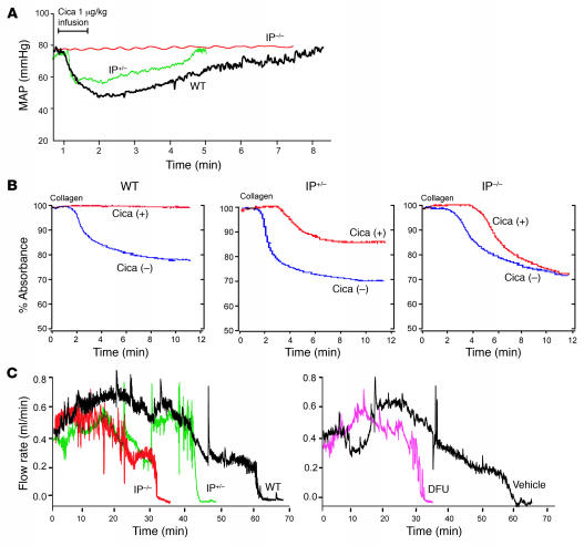
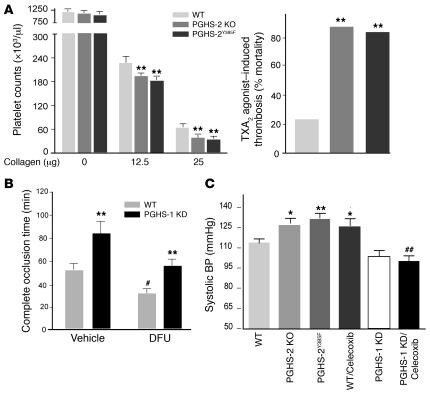
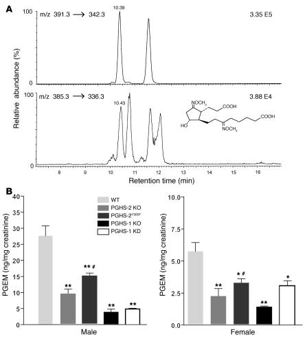
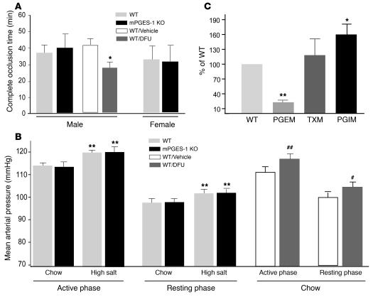
We investigated the mechanisms by which inhibitors of prostaglandin G/H synthase-2 (PGHS-2; known colloquially as COX-2) increase the incidence of myocardial infarction and stroke. These inhibitors are believed to exert both their beneficial and their adverse effects by suppression of PGHS-2-derived prostacyclin (PGI(2)) and PGE(2). Therefore, the challenge remains to identify a mechanism whereby PGI(2) and PGE(2) expression can be suppressed while avoiding adverse cardiovascular events. Here, selective inhibition, knockout, or mutation of PGHS-2, or deletion of the receptor for PGHS-2-derived PGI(2), was shown to accelerate thrombogenesis and elevate blood pressure in mice. These responses were attenuated by COX-1 knock down, which mimics the beneficial effects of low-dose aspirin. PGE(2) biosynthesis is catalyzed by the coordinate actions of COX enzymes and microsomal PGE synthase-1 (mPGES-1). We show that deletion of mPGES-1 depressed PGE(2) expression, augmented PGI(2) expression, and had no effect on thromboxane biosynthesis in vivo. Most importantly, mPGES-1 deletion affected neither thrombogenesis nor blood pressure. These results suggest that inhibitors of mPGES-1 may retain their antiinflammatory efficacy by depressing PGE(2), while avoiding the adverse cardiovascular consequences associated with PGHS-2-mediated PGI(2) suppression.


Figures

Figure 1
Figure 1. PGHS enzymes and eicosanoid biosynthesis.
(A) Urinary excretion of TXM was decreased significantly (n = 6; *P < 0.001) from values in WT mice by PGHS-1 deletion (KO) or knock down (KD), but not in PGHS-2 KO or PGHS-2Y385F mice or those treated with 100 mg/kg/d of the PGHS-2 inhibitor celecoxib for 30 days on a mixed C57BL/6 × 129/Sv genetic background. (B) Urinary PGIM was depressed significantly in PGHS-2 KO and PGHS-2Y385F mice and by celecoxib, but not in PGHS-1 KO or KD mice.
Figure 2
Figure 2. Suppressive effects of IP deletion on vascular reactivity, platelet aggregation, and thrombogenesis are gene/dose dependent.
(A) The maximum decline (P < 0.0001) and duration (P < 0.005) of hypotension evoked in mean arterial pressure (MAP) by 1 μg/kg of IP agonist, cicaprost (Cica), was greater in WT than in IP+/– or IP–/– mice on a C57BL/6 background. (B) Platelet aggregation was initiated with 2 μg/ml collagen with (+) or without (–) pretreatment with 10 nM Cica. Inhibition of aggregation was not evident in IP–/– mice and averaged 86% of WT in IP+/– mice (P < 0.0001). (C) Carotid artery blood flow after photochemical injury. The time to complete occlusion after rose bengal dye injection fell from 66.3 ± 5.1 minutes in WT to 44.4 ± 7 minutes in IP+/– and to 29.7 ± 7.6 minutes in IP–/– mice (P < 0.006). The mean impact of the COX-2 inhibitor, DFU (10 mg/kg for 3 days), on time to occlusion (56.2% of WT value) was intermediate between that of IP+/– (68.1%) and IP–/– (45.5%) mice.
Figure 3
Figure 3. PGHS-2 disruption or inhibition promotes thrombogenesis and hypertension and modulation effect of PGHS-1 KD.
(A) Circulating platelets before and 2 minutes after injection of collagen (12.5 and 25 μg/kg) plus epinephrine (15 μg/ml, 100 μl) into WT, PGHS-2Y385F, and PGHS-2 KO mice on a mixed C57BL/6 × 129/Sv background. Thrombocytopenia was more pronounced (**P < 0.01) in PGHS-2–disrupted mice. Sudden death was induced more commonly (**P < 0.01) within 15 minutes of U46619 injection in PHGS-2–deleted or –mutated mice. (B) The time to thrombotic carotid artery occlusion after photochemical injury was delayed in PGHS-1 KD mice (**P < 0.01) but accelerated by DFU treatment (#P < 0.05). The time to occlusion in DFU-treated animals was delayed in the PGHS-1 KD group compared with WT controls (**P < 0.01), while the time to occlusion in DFU-treated PGHS-1 KD mice did not differ significantly from that in vehicle-treated WT controls on a mixed C57BL/6 × 129/Sv background. (C) Systolic blood pressure, as measured by the tail cuff method, was elevated significantly in 3-month-old PGHS-2 KO, PGHSY385F, and celecoxib-treated (100 mg/kg/d for 30 days) mice as compared with WT mice on a mixed C57BL/6 × 129/Sv background (*P < 0.05; **P < 0.01). The hypertensive effect of celecoxib was attenuated in PGHS-1 KD mice compared with that in WT (##P < 0.01) mice.
Figure 4
Figure 4. The major urinary metabolite of PGE2 and the suppressive effect of PGHS-1 disruption or KD and PGHS-2 disruption or mutation on PGE2 biosynthesis.
(A) A selected ion-monitoring trace of the methoxime derivative of endogenous PGEM (9,15-dioxo-11α-hydroxy-2,3,4,5-tetranor-prostane-1,20-dioic-17,17,18,18,19,19-d6 acid) (bottom panel) and its hexadeuterated internal standard (top panel). (B) Urinary PGEM decreased significantly in both male and female PGHS-2 KO or PGHS-2Y385F mice compared with WT controls on a mixed C57BL/6 × 129/Sv genetic background (n = 5–6; *P < 0.05; **P < 0.001). PGEM was also significantly lower in PGHS-1 KD and PGHS-1 KO groups compared with WT mice of mixed C57BL/6 × 129/Sv genetic background (n = 5–6; *P < 0.05; **P < 0.001). PGEM was significantly higher in PGHS-2Y385F mice compared with PGHS-2 KO mice (#P < 0.05) on the same genetic background.
Figure 5
Figure 5. Impact of mPGES-1 disruption on thrombogenesis, blood pressure regulation, and eicosanoid biosynthesis.
(A) Deletion of mPGES-1 failed to alter the time to thrombotic carotid artery occlusion after photochemical injury, while it was accelerated by the PGHS-2 inhibitor DFU in mice on a DBA/11acJ background (*P < 0.05). (B) MAP exhibited diurnal variation in mPGES-1 KO and WT littermates on a mixed DBA/11acJ × C57BL/6 background. MAP was averaged over 4 days for 12 hours dark (active phase) and light (resting phase) periods. MAP was higher during active phase, and a high-salt diet elevated pressure similarly, a mean 6% in both groups (**P < 0.01). Oral DFU administration (10 mg/kg/d) for 21 days increased MAP in both the active (##P < 0.01) and resting (#P < 0.05) phases compared with vehicle-treated animals. (C) Urinary PGEM was lower (**P < 0.01) and PGIM was higher (*P < 0.05), while TXM was unaltered in male mPGES-1 KO mice compared with gender-matched WT littermates on a DBA/11acJ background.

References

    1. Patrignani P., et al. COX-2 is not involved in thromboxane biosynthesis by activated human platelets. J. Physiol. Pharmacol. 1999;50:661–667. - PubMed
    1. Cheng Y., et al. Role of prostacyclin in the cardiovascular response to thromboxane A2. Science. 2002;296:539–541. - PubMed
    1. FitzGerald G.A. Coxibs and cardiovascular disease. N. Engl. J. Med. 2004;351:1709–1711. - PubMed
    1. Grosser T., Fries S., FitzGerald G.A. Biological basis for the cardiovascular consequences of COX-2 inhibition: therapeutic challenges and opportunities. J. Clin. Invest. 2006;116:4–15. - PMC - PubMed
    1. McAdam B.F., et al. Systemic biosynthesis of prostacyclin by cyclooxygenase (COX)-2: the human pharmacology of a selective inhibitor of COX-2. Proc. Natl. Acad. Sci. U. S. A. 1999;96:272–277. - PMC - PubMed

Publication types