. 2006 Jun;15(6):923-9.
doi: 10.1007/s00586-005-1026-z. Epub 2006 Apr 14.

Three-dimensional finite element analysis of the pediatric lumbar spine. Part I: pathomechanism of apophyseal bony ring fracture

Affiliations

Three-dimensional finite element analysis of the pediatric lumbar spine. Part I: pathomechanism of apophyseal bony ring fracture

Koichi Sairyo et al. Eur Spine J. 2006 Jun.

Abstract

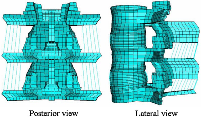
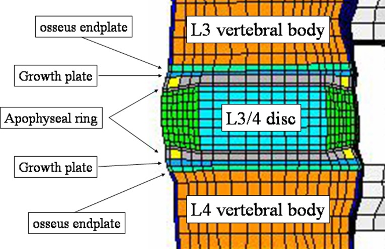
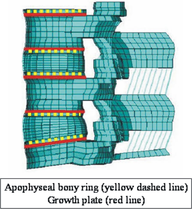
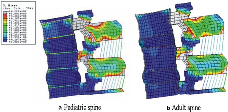
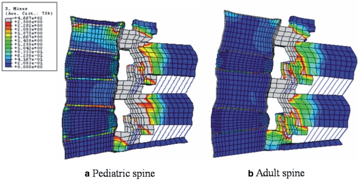
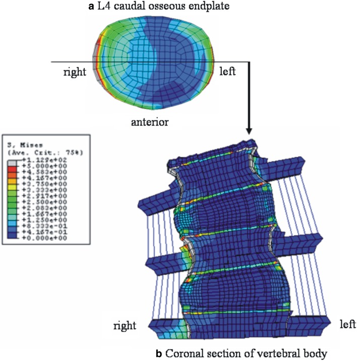
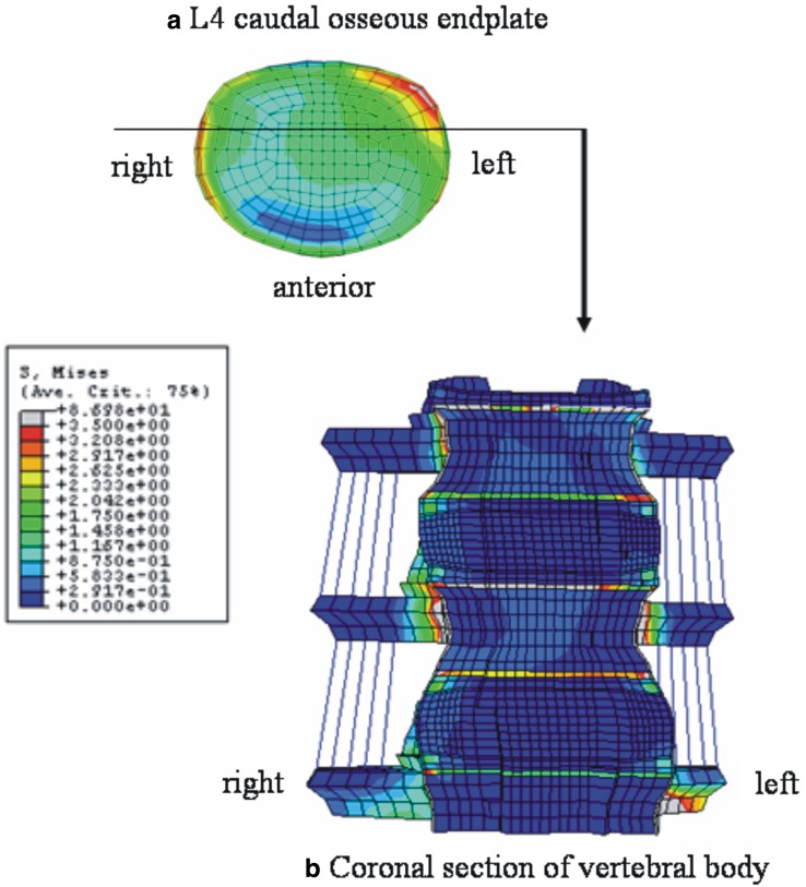
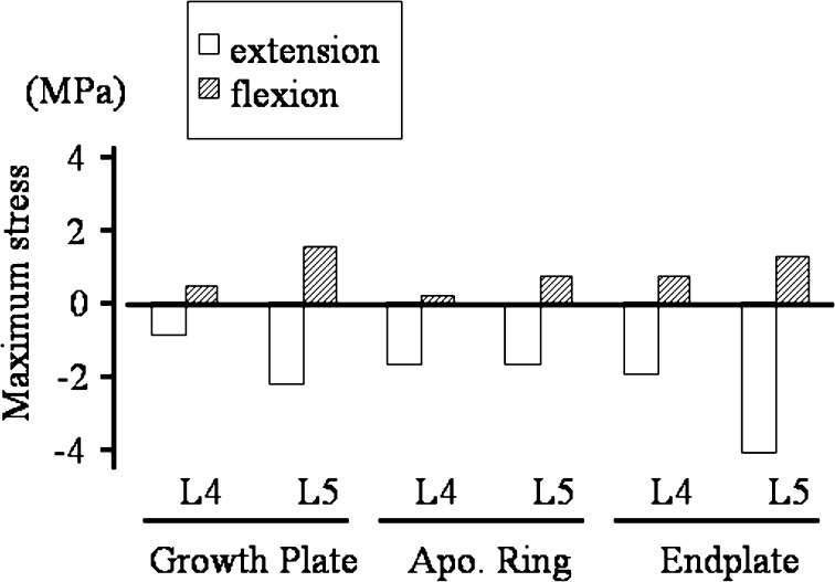
The purpose of this study was to (1) develop a three-dimensional, nonlinear pediatric lumbar spine finite element model (FEM), and (2) identify the mechanical reasons for the posterior apophyseal bony ring fracture in the pediatric patients. The pediatric spine FE model was created from an experimentally validated three-dimensional adult lumbar spine FEM. The size of the FEM was reduced to 96% taking into account of the ratio of the sitting height of an average 14-years-old children to that of an adult. The pediatric spine was created with anatomically specific features like the growth plate and the apophyseal bony ring. For the stress analyses, a 10-N m moment was applied in all the six directions of motion for the lumbar spine. A preload of 351 N was applied which corresponds to the mean body weight of the 14-years-old group. The stresses at the apophyseal bony ring, growth plate and endplate were calculated. The results indicate that the structures surrounding the growth plate including apophyseal bony ring and osseous endplate were highly stressed, as compared to other structures. Furthermore, posterior structures in extension were in compression whereas in flexion they were in tension, with magnitude of stresses higher in extension than in flexion. Over time, the higher compression stresses along with tension stresses in flexion may contribute to the apophyseal ring fracture (fatigue phenomena).


Figures

Fig. 1
Fig. 1
Experimentally validated adult L3–L5 ligamentous finite element model (FEM). The model was modified to the pediatric model
Fig. 2
Fig. 2
Sagittal section of the pediatric FEM at L3–L4. Note the layers of growth plate and apophyseal bony ring, which are the specific structures in the pediatric spine. In this model, apophyseal ring was simulated as a bony ring
Fig. 3
Fig. 3
Location of apophyseal bony ring (yellow lines) and growth plate (red lines)
Fig. 4
Fig. 4
a Stress distributions in 10 N m flexion and a preload in pediatric, and b adult spine models. In both models, higher stresses were observed around pedicle and facet joints. In the anterior structures, the stress concentration at osseous endplate and apophyseal bony ring was observed in the pediatric model, whereas, in the adult model endplate showed stress concentration
Fig. 5
Fig. 5
a Stress distributions in extension at 10 N m with precompression loading in pediatric and b adult spine models: In both models, higher stresses are observed around pedicle and facet joints during the extension motion. In the anterior structures, the stress concentration at osseous endplate and apophyseal bony ring is observed in the pediatric model, whereas, in the adult model endplate shows stress concentration
Fig. 6
Fig. 6
Stress distributions in right lateral bending at 10 N m with precompression loading in pediatric models. a Top view of the L4 caudal osseous endplate, b anterior view of the coronal section of the model through the middle of vertebral body as indicated in a. In a, both right and left lateral corner are shown to be highly stressed due to compression and traction force, respectively. The stress distribution pattern in b indicates the high loading at the osseous endplate and apophyseal bony ring
Fig. 7
Fig. 7
Stress distributions in right axial rotation at 10 N m with precompression loading in pediatric models. a Top view of the L4 caudal osseous endplate, b anterior view of the coronal section of the model through the anterior one-third of vertebral body as indicated in a. In a, left posterior–lateral corner is shown to be highly stressed. The stress distribution pattern in b indicates the high loading at the osseous endplate and apophyseal bony ring
Fig. 8
Fig. 8
Axial stresses at the posterior structures around the apophyseal ring (positive value traction, negative value compression). Posterior structures were loaded in compression in extension whereas in flexion they were in tension. The absolute values of stresses were always higher in extension than in flexion. (Apo Ring apophyseal bony ring)

Similar articles

Cited by

References

    1. Arlet V, Fassier F. Herniated nucleus pulposus and slipped vertebral apophysis. In: Weinstein SL, editor. The pediatric spine. Philadelphia: Lippincott Williams & Wilkins; 2001. pp. 453–469.
    1. Goel VK, Lim T-H, Gwon J, et al. Effects of rigidity of an internal fixation device—a comprehensive biomechanical investigation. Spine. 1991;16(Suppl):S155–S161. - PubMed
    1. Goel VK, Monroe BT, Gilbertson LG, et al. Interlaminar shear stresses and laminae separation in a disc: finite element analysis of the L3-4 motion segment subjected to axial compressive loads. Spine. 1995;20:689–698. - PubMed
    1. Goel V, Grauer J, Patel T, et al. Effects of Charite artificial disc on the implanted and adjacent spinal segments mechanics using a hybrid testing protocol. Spine. 2005;30:2755–2764. doi: 10.1097/01.brs.0000195897.17277.67. - DOI - PubMed
    1. Ikata T, Morita T, Katoh S, et al. Lesions of the lumbar posterior end plate in children and adolescents. An MRI study. J Bone Joint Surg [Br] 1995;77:951–955. - PubMed