. 2006 Apr;14(4):757-66.
doi: 10.1016/j.str.2006.01.011.

Magnesium-induced assembly of a complete DNA polymerase catalytic complex

Affiliations

Magnesium-induced assembly of a complete DNA polymerase catalytic complex

Vinod K Batra et al. Structure. 2006 Apr.

Abstract

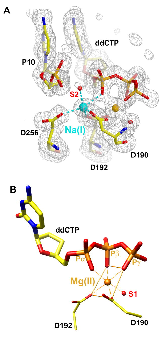
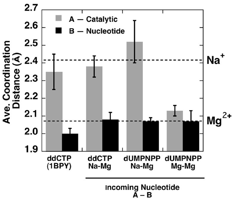
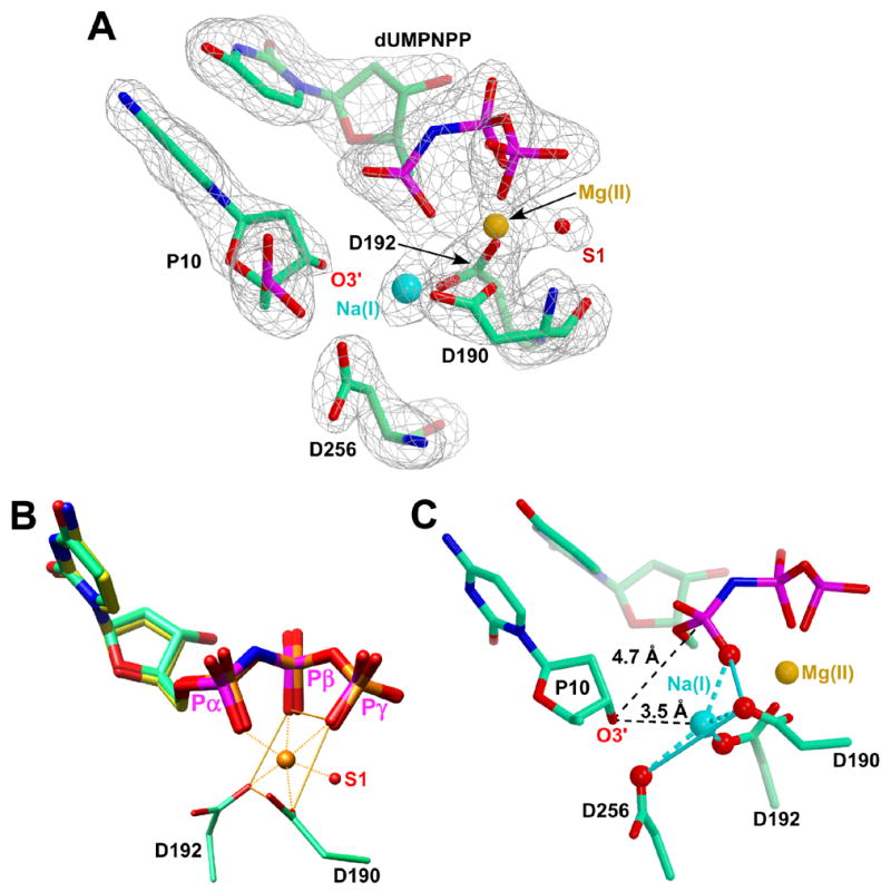
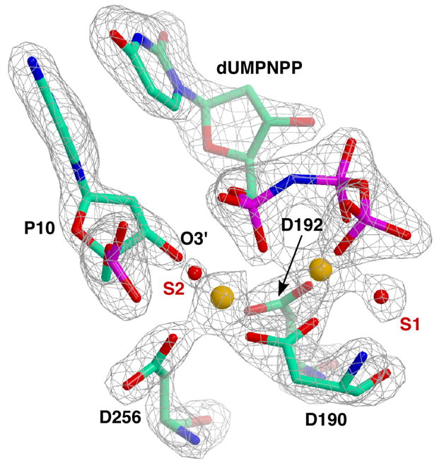
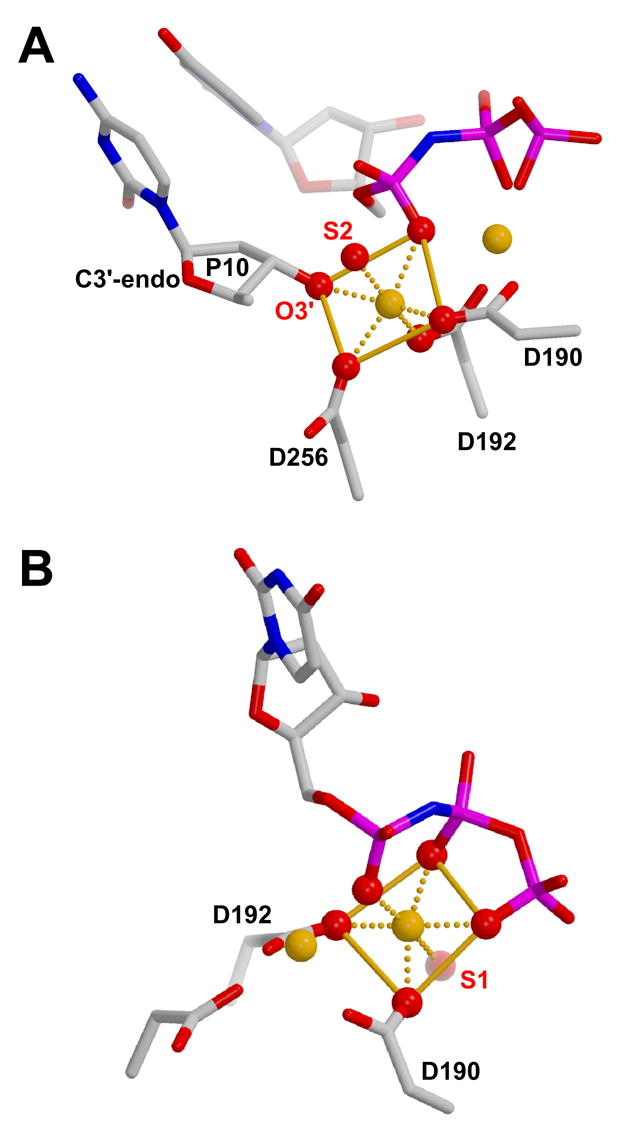
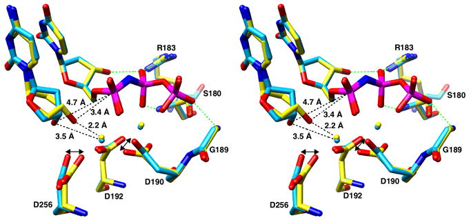
The molecular details of the nucleotidyl transferase reaction have remained speculative, as strategies to trap catalytic intermediates for structure determination utilize substrates lacking the primer terminus 3'-OH and catalytic Mg2+, resulting in an incomplete and distorted active site geometry. Since the geometric arrangement of these essential atoms will impact chemistry, structural insight into fidelity strategies has been hampered. Here, we present a crystal structure of a precatalytic complex of a DNA polymerase with bound substrates that include the primer 3'-OH and catalytic Mg2+. This catalytic intermediate was trapped with a nonhydrolyzable deoxynucleotide analog. Comparison with two new structures of DNA polymerase beta lacking the 3'-OH or catalytic Mg2+ is described. These structures provide direct evidence that both atoms are required to achieve a proper geometry necessary for an in-line nucleophilic attack of O3' on the alphaP of the incoming nucleotide.


Figures

Figure 1
Figure 1. Active Site Coordination of the Ions Observed in a High-Resolution (1.65 Å) DNA Polymerase β /DNA/ddCTP Teranry Complex
(A) A Fo-Fc simulated annealing electron density omit map (gray) contoured at 2.5σ for the pol β active site. A penta-coordinated ion is bound to the catalytic metal site. The coordination distances (Table 3) are consistent with the identity of this ion as Na+ (light blue). Additionally, a previously postulated water molecule (S2) is now observed. The Na+ is also coordinated by all three active site aspartates (D190, D192, D256) and a non-bridging oxygen on Pα (pro-RP oxygen). The nucleotide binding metal is consistent with Mg2+ (orange). The density is superimposed on the refined modeled primer terminus (P10), incoming nucleotide (ddCTP), active site aspartates (D190, D192, D256), and a nucleotide metal-coordinating water. The primer terminus O3’ is believed to be the sixth ligand, but was omitted to trap the catalytic intermediate for structure determination. (B) The octahedral coordination and coordination distances (Table 3) of the ion in the nucleotide binding metal site is consistent with the identity of this ion as Mg2+. The metal is coordinated to non-bridging oxygens on all three phosphates of the incoming ddCTP (α,β ,γ-tridentate). In addition, Asp190 (D190), Asp192 (D192), and a water molecule (S1) completes the inner coordination sphere.
Figure 2
Figure 2
Average Coordination Distances for the Oxygen Ligands to the Catalytic (A) or Nucleotide Binding (B) Metals These distances are computed for a previously reported structure of pol β (1BPY) (Sawaya et al., 1997), the 1.65 Å structure (ddCTP/Na–Mg; 2FMP), and two ternary substrate complex structures trapped with the dUMPNPP non-hydrolyzable analogue (Na–Mg, 2FMQ; Mg–Mg, 2FMS). The substrate complex structures are denoted by the identity of the incoming nucleotide and the putative ions in sites A and B. The specific coordinating atoms and distances are tabulated in Table 3. The errors bars represent the standard errors of the calculated averages. The horizontal dashed lines indicate the coordinate distances observed from accurately determined small molecule structures (Harding, 2001, Harding, 2002).
Figure 3
Figure 3. Kinetic Analysis of dUTP Insertion and dUMPNPP Inhibiiton
(A) Steady-state kinetic analysis indicates that dUTP insertion (■) is similar to that for dTTP (●). The catalytic efficiencies (kcat/Km) for dTTP and dUTP insertion are 0.19 ± 0.03 and 0.15 ± 0.02 1/μM–s, respectively, for at least two independent determinations. (B) Dixon analysis for competitive inhibition indicates that the Ki for dUMPNPP is 0.9 μM (dotted line).
Figure 4
Figure 4. Active Site Geometry of the Ternary DNA Polymerase β /DNA/dUMPNPP Complex with Na+ in the Catalytic Metal Site
(A) The Fo-Fc simulated annealing electron density omit map (gray) contoured at 3.8σ for the pol β active site with Na+ (light blue) occupying the catalytic site. The nucleotide binding metal is consistent with Mg2+ (orange). The density is superimposed on the refined modeled primer terminus (P10), incoming nucleotide analogue (dUMPNPP), active site aspartates (D190, D192, D256), and the nucleotide metal-coordinating water (S1). The density for O3’ is clearly observed but distant from the sodium ion in the catalytic metal site. (B) The ternary substrate complex structures of pol β with an incoming ddCTP (yellow carbon and orange phosphates atoms) and dUMPNPP (green carbon and magenta phosphate atoms) were superimposed with the catalytic subdomains (RMSD = 0.13 Å for 344 atom pairs). The superimposed structures clearly indicate that the bridging nitrogen (blue) between Pα and Pβ does not significantly alter the position of the phosphates. The octahedral coordination and coordination distances (Table 3) of the ion in the nucleotide binding metal site is consistent with the identity of this ion as Mg2+. (C) The Na+ (light blue) occupying the catalytic metal site displays a distorted coordination geometry. The four coordinating oxygens are displayed as red balls. The primer terminus (P10) O3’ is too distant (3.5 Å) for Na+ to participate in its coordination. Furthermore, O3’ is 4.7 Å from Pα of the incoming nucleotide analog.
Figure 5
Figure 5. Active Site Geometry of the Ternary DNA Polymerase β /DNA/dUMPNPP Complex with Mg2+ in the Catalytic Metal Site
The Fo-Fc simulated annealing electron density omit map (gray) contoured at 4.7σ for the pol β active site with Mg2+ (orange) occupying both the catalytic and nucleotide metal binding sites. The density is superimposed on the refined modeled primer terminus (P10), incoming nucleotide analogue (dUMPNPP), active site aspartates (D190, D192, D256), and the metal-coordinating waters (S1 and S2). The density for O3’ is clearly observed and the resulting C3’-endo sugar pucker positions O3’ near the catalytic metal and Pα of dUMPNPP.
Figure 6
Figure 6. Coordination Geometry of the Metal Binding Sites in the Complete Pre-Catalytic Complex
(A) Coordination geometry of Mg2+ (orange) occupying the catalytic metal site. The sugar pucker of the primer terminus (P10) is C3’-endo and the catalytic metal exhibits good octahedral geometry. Additionally, a water molecule (S2) not observed in the lower resolution Na+ structure is visible and participates in the first coordination sphere of the catalytic Mg2+. (B) The Mg2+ occupying the nucleotide metal site also exhibits good octahedral geometry even when the catalytic site is occupied by Na+ (Figure 4B).
Figure 7
Figure 7. Stereo View of the Pol β Active Site
Superimposed structures of pol β with either Na+ (light blue) or Mg2+ (yellow) in the catalytic metal site A. Residues that hydrogen bond (green) to the triphosphate moiety of the incoming nucleotide and the active site aspartates are shown. When Na+ occupies the catalytic metal site, O3’ of the primer terminus is 3.5 and 4.7 Å from the catalytic metal and αP of the incoming nucleotide, respectively. In contrast, with Mg2+ in the catalytic metal site, an altered sugar pucker positions O3’ of the primer terminus 2.2 and 3.4 Å from the catalytic metal and Pα of the incoming nucleotide, respectively.

Similar articles

Cited by

References

    1. Arndt JW, Gong W, Zhong X, Showalter AK, Liu J, Dunlap CA, Lin Z, Paxson C, Tsai MD, Chan MK. Insight into the catalytic mechanism of DNA polymerase β: Structures of intermediate complexes. Biochemistry. 2001;40:5368–5375. - PubMed
    1. Batra VK, Beard WA, Shock DD, Pedersen LC, Wilson SH. Nucleotide-induced DNA polymerase active site motions accomodating a mutagenic DNA intermediate. Structure (Camb) 2005;13:1225–1233. - PubMed
    1. Beard WA, Wilson SH. Purification and domain-mapping of mammalian DNA polymerase β. Methods Enzymol. 1995;262:98–107. - PubMed
    1. Beard WA, Wilson SH. Structural insights into DNA polymerase β fidelity: Hold tight if you want it right. Chem Biol. 1998;5:R7–R13. - PubMed
    1. Beard WA, Wilson SH. Structural design of a eukaryotic DNA repair polymerase: DNA polymerase β. Mutat Res. 2000;460:231–244. - PubMed

Publication types