

Review
doi: 10.1016/j.bbalip.2006.02.020.
Epub 2006 Mar 24.
Membrane binding domains
Affiliations
- PMID: 16616874
- PMCID: PMC2049088
- DOI: 10.1016/j.bbalip.2006.02.020
Item in Clipboard
Review
Membrane binding domains
Biochim Biophys Acta.
2006 Aug.
Abstract
Eukaryotic signaling and trafficking proteins are rich in modular domains that bind cell membranes. These binding events are tightly regulated in space and time. The structural, biochemical, and biophysical mechanisms for targeting have been worked out for many families of membrane binding domains. This review takes a comparative view of seven major classes of membrane binding domains, the C1, C2, PH, FYVE, PX, ENTH, and BAR domains. These domains use a combination of specific headgroup interactions, hydrophobic membrane penetration, electrostatic surface interactions, and shape complementarity to bind to specific subcellular membranes.
Figures
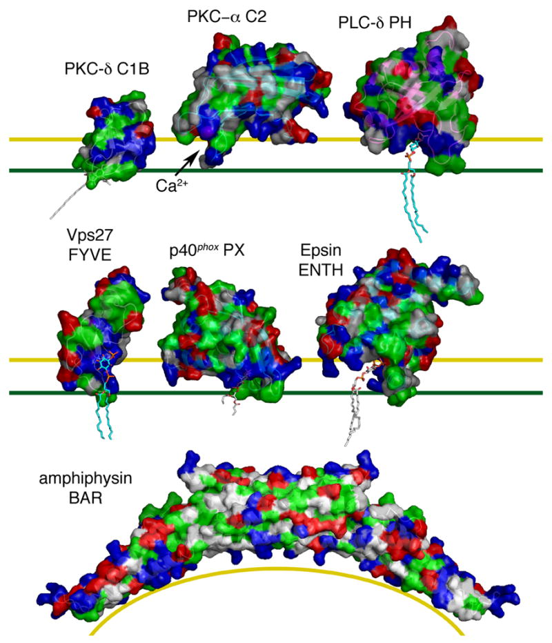
Surfaces are rendered with hydrophobic residues in green, basic in blue, acidic in red, and uncharged polar in white. The yellow line marks the top of the polar interface region of the bilayer, and the green line marks the top of the hydrocarbon core of the bilayer. The C1B domain of PKC-δ bound to phorbol 13-acetate (1PTR) [11] was docked to the membrane on the basis of structural modeling and solution NMR of the related PKC-γ C1B domain [27]. The C2 domain of PKC-α (1DSY) [39] was docked on the basis of FRET and EPR analysis [78]. The PH domain of PLC-δ (1MAI) [13] was docked on the basis of structural modeling. The FYVE domain of Vps27 (1VFY) [56], with PI(3)P docked on the basis of its binding site in EEA1 [58], was docked to the membrane based on structural and electrostatic modeling [74]. The PX domain of p40phox bound to PI(3)P (1H6H) [62] was docked on the basis of structural modeling and demonstrated membrane penetration [77]. The ENTH domain of epsin (1HOA) [63] bound to PI(4,5)P2 was docked on the basis of structural modeling and demonstrated membrane penetration [79]. The amphiphysin BAR domain (1URU) [68] was docked to a curved membrane surface based on shape complementarity. Phospholipid and phorbol ester tail moieties were modeled arbitrarily except for the PX domain, where the crystallized conformation is shown.
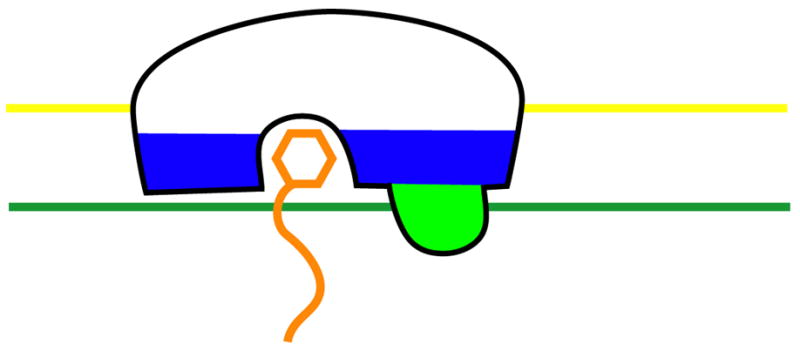
An idealized membrane binding domain is shown in the same color scheme used in Fig. 1.
References
-
- Quest AFG, Bardes ESG, Bell RM. A phorbol ester binding domain of protein kinase-C-γ-high-affinity binding to a glutathione-S-transferase/cys2 fusion protein. J Biol Chem. 1994;269:2953–2960. - PubMed
-
- Kazanietz MG, Bustelo XR, Barbacid M, Kolch W, Mischak H, Wong G, Pettit GR, Bruns JD, Blumberg PM. Zinc-finger domains and phorbol ester pharmacophore - analysis of binding to mutated form of protein kinase-c-τ and the vav and c-raf protooncogene products. J Biol Chem. 1994;269:11590–11594. - PubMed
-
- Davletov BA, Sudhof TC. A single C2 domain from synaptotagmin-I is sufficient for high-affinity Ca2+/phospholipid binding. J Biol Chem. 1993;268:26386–26390. - PubMed
-
- Haslam RJ, Koide HB, Hemmings BA. Pleckstrin domain homology. Nature. 1993;363:309–310. - PubMed
Publication types
MeSH terms
Substances
Grants and funding
LinkOut - more resources
Full Text Sources
Other Literature Sources
Miscellaneous
