. 2006 Apr 25;103(17):6506-11.
doi: 10.1073/pnas.0601212103. Epub 2006 Apr 14.

EM measurements define the dimensions of the "30-nm" chromatin fiber: evidence for a compact, interdigitated structure

Affiliations

EM measurements define the dimensions of the "30-nm" chromatin fiber: evidence for a compact, interdigitated structure

Philip J J Robinson et al. Proc Natl Acad Sci U S A. .

Abstract

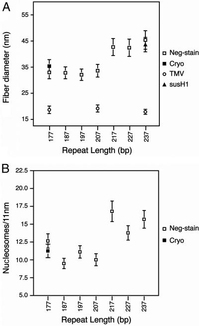
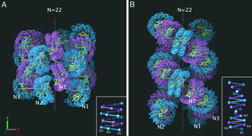
Chromatin structure plays a fundamental role in the regulation of nuclear processes such as DNA transcription, replication, recombination, and repair. Despite considerable efforts during three decades, the structure of the 30-nm chromatin fiber remains controversial. To define fiber dimensions accurately, we have produced very long and regularly folded 30-nm fibers from in vitro reconstituted nucleosome arrays containing the linker histone and with increasing nucleosome repeat lengths (10 to 70 bp of linker DNA). EM measurements show that the dimensions of these fully folded fibers do not increase linearly with increasing linker length, a finding that is inconsistent with two-start helix models. Instead, we find that there are two distinct classes of fiber structure, both with unexpectedly high nucleosome density: arrays with 10 to 40 bp of linker DNA all produce fibers with a diameter of 33 nm and 11 nucleosomes per 11 nm, whereas arrays with 50 to 70 bp of linker DNA all produce 44-nm-wide fibers with 15 nucleosomes per 11 nm. Using the physical constraints imposed by these measurements, we have built a model in which tight nucleosome packing is achieved through the interdigitation of nucleosomes from adjacent helical gyres. Importantly, the model closely matches raw image projections of folded chromatin arrays recorded in the solution state by using electron cryo-microscopy.


Conflict of interest statement

Conflict of interest statement: No conflicts declared.

Figures

Fig. 1.
Fig. 1.
Relationship between linker length and chromatin fiber diameter. (A) Gallery of side views of negatively stained fibers of 72 × 177-bp (177), 52 × 187-bp (187), 61 × 197-bp (197), 47 × 207-bp (207), 55 × 217-bp (217), 66 × 227-bp (227), and 56 × 237-bp (237) 601 nucleosome arrays reconstituted with saturating concentrations of histone octamer and linker histone H5 and folded in 1.6 mM MgCl2. Images show both individual fibers and longer aggregates formed through the end-to-end stacking of folded arrays as described for native chromatin containing linker histone (48, 53). (B) Images of the 72 × 177-bp fibers captured in the frozen hydrated state by using electron cryo-microscopy. (Scale bar: 100 nm.)
Fig. 2.
Fig. 2.
Plots of the diameter and length of the reconstituted and folded 601-nucleosome arrays shown in Fig. 1. (A) Relationship between fiber diameter and linker length. The average diameter dimensions shown are calculated from 150–300 measurements of negatively stained (open squares) and frozen hydrated (filled square) chromatin arrays folded in 1.6 mM MgCl2. Average diameters of TMV (open circles) and the 56 × 237-bp 601 array reconstituted with linker histone H1 purified from sea urchin sperm (filled triangle) are also included. (B) Relationship between the number of nucleosomes and chromatin fiber length. The average length dimensions shown are calculated from 70 to 100 measurements of negatively stained (open squares) and frozen hydrated (filled square) chromatin arrays folded in 1.6 mM MgCl2.
Fig. 3.
Fig. 3.
Modeling the 30-nm chromatin fiber. (A) Interdigitated one-start helix model built by using the constraints imposed by our measurements of diameter and nucleosome packing ratio (Fig. 2). The helix contains 22 nucleosomes and has a diameter of 33 nm and height of ≈33 nm. Alternate helical gyres are colored marine and magenta. (B) Two-start helical crossed linker model adapted from the idealized model reported by Schalch et al. (38). The model maintains the same parameters for rise per nucleosome and azimuthal rotation angle while extrapolating to a 177-bp nucleosome repeat length by increasing the radius by 3.4 nm (roughly approximating the addition of 10 bp of extra linker DNA lying orthogonal to the fiber axis). The resulting helix contains 22 nucleosomes and has a 28.4-nm diameter and ≈47-nm height. Alternate nucleosome pairs are colored marine and magenta. The positions of the first, second, third, and seventh nucleosomes in the linear DNA sequence are marked on both models with N1, N2, N3, and N7. (Insets) Schematic representations of both atomic models showing the proposed DNA connectivity.
Fig. 4.
Fig. 4.
Comparison of models to raw images of folded chromatin. (A) Electron cryo-microscopy images of a folded chromatin array containing 22 nucleosomes with a repeat length of 177 bp. The particles are visualized unperturbed in the frozen hydrated state and oriented randomly in ice containing 1.0 mM Mg2+. The particles in each row represent similar views. (Scale bar: 100 nm.) (B) Views of the interdigitated one-start helix model (Fig. 3A) that closely match the EM images from the corresponding row in A. (C) Views of the adapted two-start crossed-linker model (Fig. 3B) that are equivalent to those of the interdigitated model from the corresponding row in B. The varying orientation of the coordinate axes describes the three-dimensional orientation of the models in the corresponding rows.

References

    1. Davies H. G., Small J. V. Nature. 1968;217:1122–1125. - PubMed
    1. Ris H., Kubai D. F. Annu. Rev. Genet. 1970;4:263–294. - PubMed
    1. Finch J. T., Lutter L. C., Rhodes D., Brown R. S., Rushton B., Levitt M., Klug A. Nature. 1977;269:29–36. - PubMed
    1. Richmond T. J., Finch J. T., Rushton B., Rhodes D., Klug A. Nature. 1984;311:532–537. - PubMed
    1. Luger K., Mader A. W., Richmond R. K., Sargent D. F., Richmond T. J. Nature. 1997;389:251–260. - PubMed

Publication types