. 2006 Apr 25;103(17):6641-6.
doi: 10.1073/pnas.0600743103. Epub 2006 Apr 14.

Disparate thermodynamics governing T cell receptor-MHC-I interactions implicate extrinsic factors in guiding MHC restriction

Affiliations

Disparate thermodynamics governing T cell receptor-MHC-I interactions implicate extrinsic factors in guiding MHC restriction

Lauren K Ely et al. Proc Natl Acad Sci U S A. .

Abstract

The underlying basis of major histocompatibility complex (MHC) restriction is unclear. Nevertheless, current data suggest that a common thermodynamic signature dictates alphabeta T cell receptor (TcR) ligation. To evaluate whether this thermodynamic signature defines MHC restriction, we have examined the thermodynamic basis of a highly characterized immunodominant TcR interacting with its cognate peptide-MHC-I ligand. Surprisingly, we observed this interaction to be governed by favorable enthalpic and entropic forces, which is in contrast to the prevailing generality, namely, enthalpically driven interactions combined with markedly unfavorable entropic forces. We conclude that extrinsic molecular factors, such as coreceptor ligation, conformational adjustments involved in TcR signaling, or constraints dictated by higher-order arrangement of ligated TcRs, might play a greater role in guiding MHC restriction than appreciated previously.


Conflict of interest statement

Conflict of interest statement: No conflicts declared.

Figures
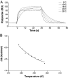
Fig. 1.
Fig. 1.
Thermodynamic analysis of the interaction between LC13 TcR and FLR–HLA-B8. (A) Temperature dependence of the LC13 TcR-binding FLR–HLA-B8 detected by SPR. RU, resonance units. (B) Free energy (ΔG) values over the temperature range of 5–25°C. Each data point is the mean of at least two independent SPR experiments; nonlinear regression was used to fit the three-parameter equation to calculate ΔH, ΔS, and ΔCp.
Fig. 2.
Fig. 2.
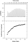
The change in enthalpy at binding was measured directly by ITC. (Upper) Heat released over time for the titration of LC13 TcR into FLR–HLA-B8 at 25°C. (Lower) Baseline-subtracted data expressed as the molar ratio plotted against the area under the peak and fitted by a model for single-site binding.
Fig. 3.
Fig. 3.
The LC13 TcR forms an extensive hydrogen-bonding network stabilizing the CDR loops in the unliganded conformation. The binding interface of the unliganded TcR is depicted with the side chains of the residues that form inter-CDR contacts and the hydrogen-bonding network that they form (gray dashed lines).
Fig. 4.
Fig. 4.
Hydrophobic regions of the binding interface correspond to the energetic hot spot of binding. (A) Surface representation of the LC13 TcR binding interface highlighting the hydrophobic (green) and hydrophilic (blue) atomic contacts with FLR–HLA-B8. The FLR peptide and HLA-B8 helices (yellow) have been overlaid to indicate the binding orientation. (B) Residues that interact with FLR–HLA-B8 have been colored according to the impact they have on binding as determined by alanine-scanning mutagenesis (18). Residues colored red have the most significant impact, followed by shades of yellow, green, and blue; dark blue residues have no effect on binding.
Fig. 5.
Fig. 5.
During ligation the LC13 TcR expels solvent from the binding interface. (A) Surface representation (mesh) of the unliganded LC13 TcR. Water molecules associated with the residues that form the binding interface at ligation and those that form bridging H bonds between the α (orange) and β (yellow) chains of the TcR are shown as cyan spheres (17). (B) Ligated conformation of the LC13 TcR bound to the FLR–HLA-B8 complex (purple). Water molecules that form bridging H bonds between the TcR and FLR–HLA-B8 are shown in blue, whereas those that remain associated with only the TcR are depicted in cyan (10).

References

    1. van der Merwe P. A., Davis S. J. Annu. Rev. Immunol. 2003;21:659–684. - PubMed
    1. Rudolph M. G., Wilson I. A. Curr. Opin. Immunol. 2002;14:52–65. - PubMed
    1. Ely L. K., Kjer-Nielsen L., McCluskey J., Rossjohn J. IUBMB Life. 2005;57:575–582. - PubMed
    1. Garcia K. C., Adams E. J. Cell. 2005;122:333–336. - PubMed
    1. Wu L. C., Tuot D. S., Lyons D. S., Garcia K. C., Davis M. M. Nature. 2002;418:552–556. - PubMed

Publication types

MeSH terms