. 2006 Apr 25;45(16):5158-70.
doi: 10.1021/bi0517887.

Semisynthesis and characterization of mammalian thioredoxin reductase

Affiliations

Semisynthesis and characterization of mammalian thioredoxin reductase

Brian Eckenroth et al. Biochemistry. .

Abstract

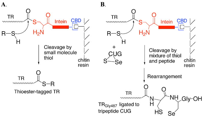
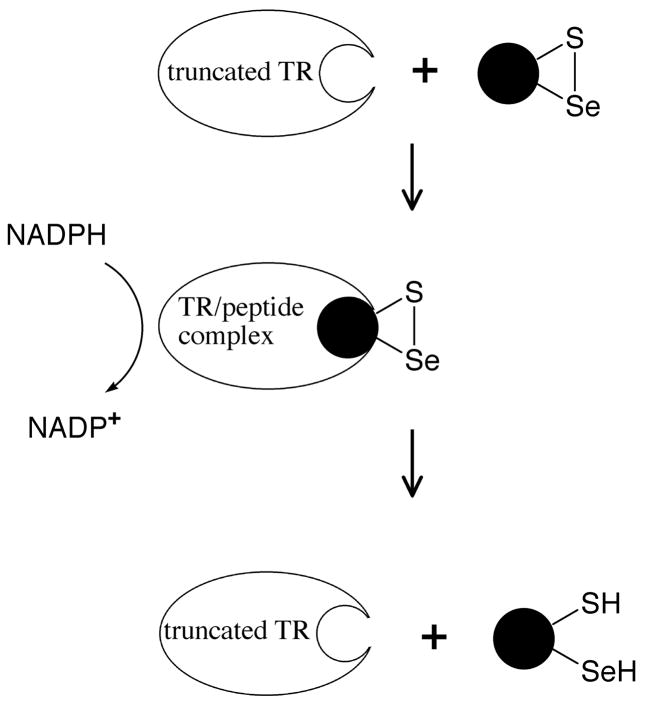
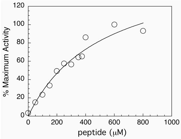
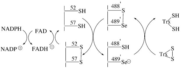
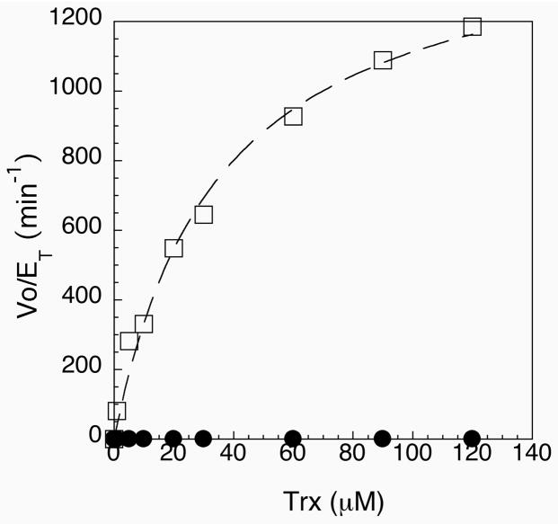
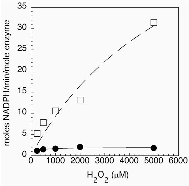
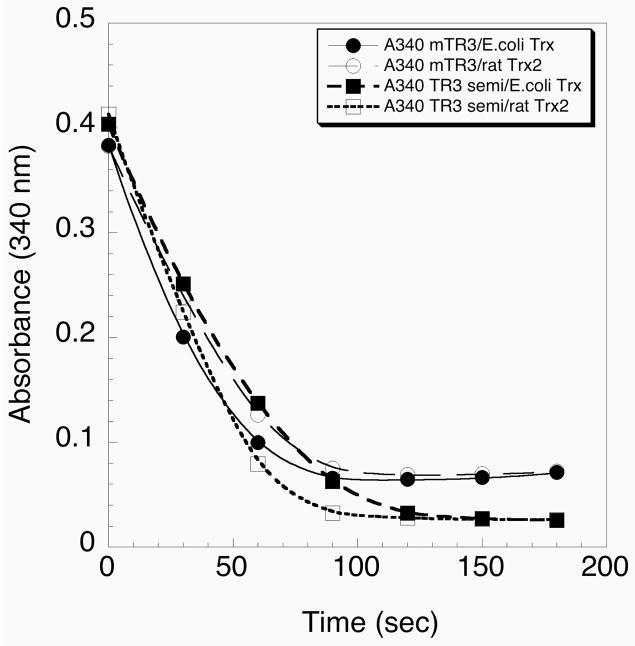
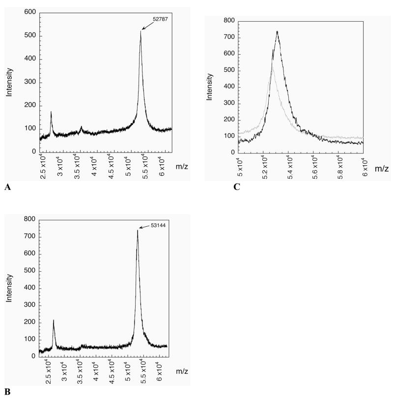
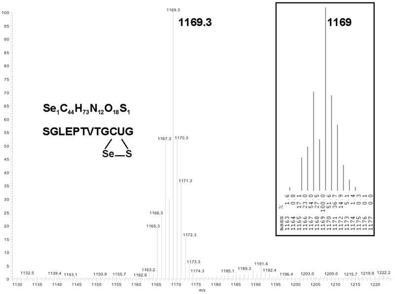
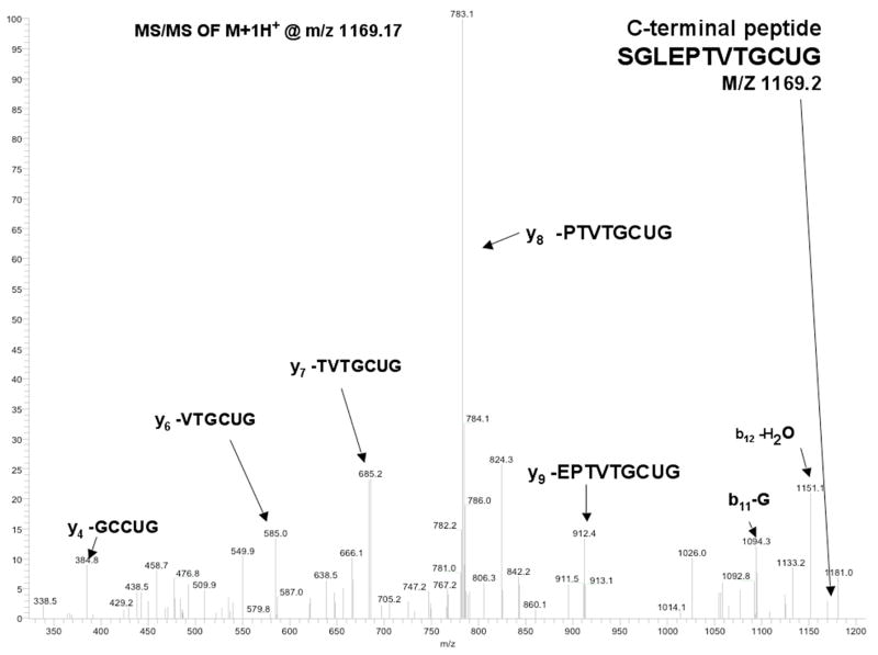
Thioredoxin reductase and thioredoxin constitute the cellular thioredoxin system, which provides reducing equivalents to numerous intracellular target disulfides. Mammalian thioredoxin reductase contains the rare amino acid selenocysteine. Known as the "21st" amino acid, selenocysteine is inserted into proteins by recoding UGA stop codons. Some model eukaryotic organisms lack the ability to insert selenocysteine, and prokaryotes have a recoding apparatus different from that of eukaryotes, thus making heterologous expression of mammalian selenoproteins difficult. Here, we present a semisynthetic method for preparing mammalian thioredoxin reductase. This method produces the first 487 amino acids of mouse thioredoxin reductase-3 as an intein fusion protein in Escherichia coli cells. The missing C-terminal tripeptide containing selenocysteine is then ligated to the thioester-tagged protein by expressed protein ligation. The semisynthetic version of thioredoxin reductase that we produce in this manner has k(cat) values ranging from 1500 to 2220 min(-)(1) toward thioredoxin and has strong peroxidase activity, indicating a functional form of the enzyme. We produced the semisynthetic thioredoxin reductase with a total yield of 24 mg from 6 L of E. coli culture (4 mg/L). This method allows production of a fully functional, semisynthetic selenoenzyme that is amenable to structure-function studies. A second semisynthetic system is also reported that makes use of peptide complementation to produce a partially active enzyme. The results of our peptide complementation studies reveal that a tetrapeptide that cannot ligate to the enzyme (Ac-Gly-Cys-Sec-Gly) can form a noncovalent complex with the truncated enzyme to form a weak complex. This noncovalent peptide-enzyme complex has 350-500-fold lower activity than the semisynthetic enzyme produced by peptide ligation.


Figures

Figure 1
Figure 1
(A) Thiol-mediated cleavage of mTR-3-intein fusion construct by addition of exogenous thiol. The junction between mTR-3 and the intein exists in equilibrium between amide and thioester forms. Addition of exogenous thiol causes cleavage of the target protein from the thioester form of the fusion protein. (B) Mechanism of incorporation of tripeptide CUG to mTR-3-intein fusion protein. The ligation is done by adding oxidized tripeptide to the cleavage buffer containing 70 mM thiol. The excess thiol both reduces the tripeptide and causes cleavage of mTR-3 from the intein fusion. The reduced peptide can then attack the thioester-tagged protein and become stably ligated to the protein because of the presence of the N-terminal amino group which enables rapid rearrangement to the amide form of the protein.
Figure 2
Figure 2
Peptide complementation to create a transient-semisynthetic TR. Oxidized peptides containing selenocysteine can serve as substrates for the truncated enzyme because they fit into the active site of the enzyme and are able to be reduced by the N-terminal dithiol (Figure 6). This system is unlike the S-protein/S-peptide system because the peptide binds weakly to the active site. For simplicity, the N-terminal redox-active disulfide and the other subunit of the TR dimer is not shown.
Figure 3
Figure 3
(A) A 12% SDS-PAGE gel showing the effect of addition of thiol to the TR-intein fusion protein. The fusion protein was isolated in a semi-pure form by DEAE ion exchange chromatography as the first step instead of the chitin affinity resin. Fractions containing the fusion protein were then pooled. The protein sample was then loaded onto the 12% gel with or without 50 mM DTT. Lane 1: molecular weight markers. Lane 2: (10 μL loading) TR-intein fusion protein without addition of DTT. The fusion protein appears as a band near 110 kDa. Some hydrolysis of the fusion protein is apparent. Very high molecular weight bands are also present, most likely due to disulfide cross-linking in the absence of reducing agent. Lane 3: (10 μL loading) TR-intein fusion protein sample treated with 50 mM DTT. A very large band at 55 kDa now appears. Lane 4: Blank. Lane 5: the sample as in lane 2 except that 5 μL was loaded. Lane 6: same as lane 3 except that 5 μL was loaded. (B) 12% SDS-PAGE characterization of recombinant mouse thioredoxin reductase 3. Lane 1: Cell culture pre-induction. Lane 2: IPTG-induced cell culture. Lane 3: Supernatant of the cell lysate. Lane 4 and 8: Molecular weight markers. Lane 5: Truncated TR cleaved from the intein on-resin by 2-mercaptoethanol and eluted from the chitin resin. Lane 6: Truncated TR eluted from the phenyl sepharose column. Lane 7: Truncated TR after elution from the DEAE column. Lane 9: The purified semisynthetic TR after elution from the DEAE column. Lane 10: Purified truncated TR cleaved from the intein on-resin by the amino acid cysteine.
Figure 4
Figure 4
Assay of peptide complementation experiment using truncated mTR-3 and synthetic peptide Ac-GCUG. The consumption of NADPH is monitored at 340 nm. When no peptide is added to the cuvette, no decrease in absorbance is observed. However, as increasing amounts of peptide are added to the assay, a progressive consumption of NADPH is observed.
Figure 5
Figure 5
Saturation curve for the addition of tetrapeptide Ac-GCUG to the truncated enzyme. The enzyme becomes saturated at concentrations above 1 mM, thus the association between the peptide and truncated enzyme is weak.
Figure 6
Figure 6
Proposed reaction mechanism for catalysis by mouse thioredoxin reductase-3. The “prime” designation for residues 488 and 489 indicate that they are present on the other subunit of the TR dimer.
Figure 7
Figure 7
Michaelis-Menten plot of Vo/ET vs. thioredoxin concentration for semisynthetic enzyme (open squares) and the Sec489Cys mutant (closed circles). Both curves could be fitted to a hyperbolic plot. Values of kcat and Km were obtained from the curve fit and are listed in Table 2.
Figure 8
Figure 8
Peroxidase activity of semisynthetic enzyme (open squares) and the Sec489Cys mutant (closed circles). Activity is reported as moles of NADPH oxidized per minute per mole of enzyme.
Figure 9
Figure 9
Comparison of the activities of semisynthetic TR and recombinant TR. The activity of the recombinant TR produced in E. coli by using an engineered SECIS element is represented by the circles. The activity of the semisynthetic TR is represented by the squares. E. coli thioredoxin (closed symbol) and rat thioredoxin-2 (open symbol) were used as substrates.
Figure 10
Figure 10
(A) MALDI-MS of truncated mTR-3 missing the C-terminal tripeptide. The average mass for the carboxylic acid form of the enzyme is 52,857 and the mass of the thioester-tagged enzyme is 52,944. The mass difference between the two forms is 87 daltons which makes identifying the exact form of the enzyme impossible at the resolution of the spectrometer (1/100). The observed mass of 52,787 is closer to the carboxylate form of the enzyme. (B) MALDI-MS of the semisynthetic form of mTR-3. The observed mass of 53,144 is in good agreement with the predicted mass of 53,164 (oxidized form). The increase in mass between the two forms is 357 daltons, in good agreement with the expected mass increase of 329 daltons. (C) Overlay of the two spectra. The semisynthetic spectrum is in black and the truncated spectrum is in light gray. The difference in mass is clearly observable. The semisynthetic spectrum has a shoulder which lines up with the peak of the truncated spectrum. The peak width of the semisynthetic enzyme shows that it is a mixture of the two forms.
Figure 11
Figure 11
ESI-MS of peptide of sequence SGLEPTVTGCUG. The peptide corresponds to the C-terminal peptide of mTR-3 containing the ligated tripeptide CUG. The inset at right is the predicted isotope pattern for this peptide containing selenium. The mass of the peptide corresponds to the oxidized form as would be expected for a peptide containing selenocysteine.
Figure 12
Figure 12
Legend: MS/MS of peptide SGLEPTVTGCUG. The peptide was fragmented by CID. Ions produced by fragmentation are labeled in the figure with the corresponding peptide sequence.

References

    1. Mustacich D, Powis G. Thioredoxin reductase. Biochem J. 2000;346:1–8. - PMC - PubMed
    1. Holmgren A. Thioredoxin and glutaredoxin systems. J Biol Chem. 1989;264:13963–13966. - PubMed
    1. Arnér ES, Holmgren A. Physiological functions of thioredoxin and thioredoxin reductase. Eur J Biochem. 2000;267:6102–6109. - PubMed
    1. Atkins JF, Gesteland RF. The twenty-first amino acid. Nature. 2000;407:463–465. - PubMed
    1. Bock A, Forchhammer K, Heider J, Leinfelder W, Sawers G, Veprek B, Zinoni F. Selenocysteine: the 21st amino acid. Mol Microbiol. 1991;5:515–520. - PubMed

Publication types

MeSH terms