. 2006 Apr 25;45(16):5217-27.
doi: 10.1021/bi052062d.

Solution structures of spinach acyl carrier protein with decanoate and stearate

Affiliations

Solution structures of spinach acyl carrier protein with decanoate and stearate

Gregory A Zornetzer et al. Biochemistry. .

Abstract

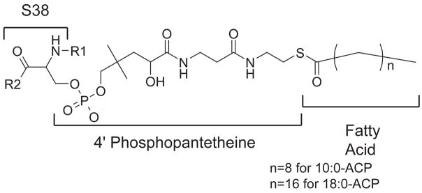
Acyl carrier protein (ACP) is a cofactor in a variety of biosynthetic pathways, including fatty acid metabolism. Thus, it is of interest to determine structures of physiologically relevant ACP-fatty acid complexes. We report here the NMR solution structures of spinach ACP with decanoate (10:0-ACP) and stearate (18:0-ACP) attached to the 4'-phosphopantetheine prosthetic group. The protein in the fatty acid complexes adopts a single conformer, unlike apo- and holo-ACP, which interconvert in solution between two major conformers. The protein component of both 10:0- and 18:0-ACP adopts the four-helix bundle topology characteristic of ACP, and a fatty acid binding cavity was identified in both structures. Portions of the protein close in space to the fatty acid and the 4'-phosphopantetheine were identified using filtered/edited NOESY experiments. A docking protocol was used to generate protein structures containing bound fatty acid for 10:0- and 18:0-ACP. In both cases, the predominant structure contained fatty acid bound down the center of the helical bundle, in agreement with the location of the fatty acid binding pockets. These structures demonstrate the conformational flexibility of spinach ACP and suggest how the protein changes to accommodate its myriad binding partners.


Figures

Figure 1
Figure 1
Covalent structure of the acyl phosphopantetheine modification of Ser38 in spinach ACP.
Figure 2
Figure 2
15N HSQC spectra of holo- and 10:0-ACP. Holo-ACP is shown in red, and 10:0-ACP is shown in blue. Samples contained 2 mM protein in 100 mM NaCl, and 10 mM MES. The pH was 6.1 and the sample temperature was 14°C. Data were collected on a Bruker DMX 500 MHz spectrometer.
Figure 3
Figure 3
Two orientations of the ensemble of 20 conformers representing the structure of 10:0-ACP with docked decanoate. The protein backbone Cα trace is shown in rainbow colors (blue N-terminus, red C-terminus). The ensemble of docked fatty acids is shown as beige sticks.
Figure 4
Figure 4
Two orientations of the ensemble of 20 conformers representing the structure of 18:0-ACP with docked stearate. The protein backbone Cα trace is shown in rainbow colors (blue N-terminus, red C-terminus). The ensemble of docked stearate is shown in beige sticks.
Figure 5
Figure 5
Two orientations showing the phosphopantetheine localization in 10:0-ACP (a) and 18:0-ACP (b). The ensemble of phosphopantetheine structures is shown as a white surface. The protein is shown as a rainbow ribbon (blue N-terminus, red C-terminus) Carbon and nitrogen (black and blue, respectively) atoms of the protein exhibiting NOE contacts to the phosphopantetheine in 10:0-ACP are shown as spheres.
Figure 6
Figure 6
Fatty acid binding pocket. Residues identified by CastP (39, 40) as lining the fatty acid binding pocket are shown as a green surface. The position of the fatty acid is shown as a beige surface enclosing a ball-and-stick representation of the fatty acid. The protein is shown as a rainbow ribbon (blue N-terminus, red C-terminus). (a) The crystal structure of E. coli butyryl-ACP (17), (b) one structure of 10:0-ACP, and (c) one structure of 18:0-ACP.

Similar articles

Cited by

References

    1. Magnuson K, Jackowski S, Rock CO, Cronan JE., Jr Regulation of fatty acid biosynthesis in Escherichia coli. Microbiol Rev. 1993;57:522–542. - PMC - PubMed
    1. Issartel J, Koronakis V, Hughes C. Activation of Escherichia-coli Prohaemolysin to the Mature Toxin by Acyl Carrier Protein-Dependent Fatty Acylation. Nature. 1991;351:759–761. - PubMed
    1. Rumley M, Therisod H, Weissborn A, Kennedy E. Mechanisms of Regulation of the Biosynthesis of Membrane-Derived Oligosaccharides in Escherichia coli. J Biol Chem. 1992;267:11806–11810. - PubMed
    1. Therisod H, Weissborn A, Kennedy E. An Essential Function for Acyl Carrier Protein in the Biosynthesis of Membrane-Derived Oligosaccharides of Escherichia coli. Proc Natl Acad Sci. 1986;83:7236–7240. - PMC - PubMed
    1. Shen B, Summers RG, Gramajo H, Bibb MJ, Hutchinson CR. Purification and characterization of the acyl carrier protein of the Streptomyces glaucescens tetracenomycin C polyketide synthase. J Bacteriol. 1992;174:3818–3821. - PMC - PubMed

Publication types