. 2006 May 15;203(5):1221-33.
doi: 10.1084/jem.20051772. Epub 2006 Apr 17.

Platelets secrete stromal cell-derived factor 1alpha and recruit bone marrow-derived progenitor cells to arterial thrombi in vivo

Affiliations

Platelets secrete stromal cell-derived factor 1alpha and recruit bone marrow-derived progenitor cells to arterial thrombi in vivo

Steffen Massberg et al. J Exp Med. .

Abstract

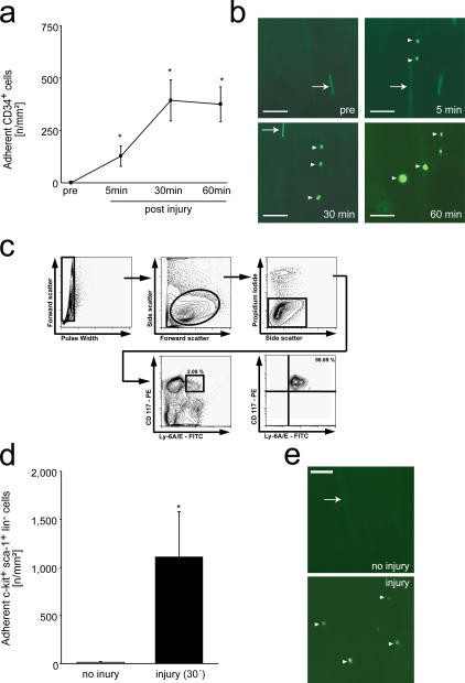
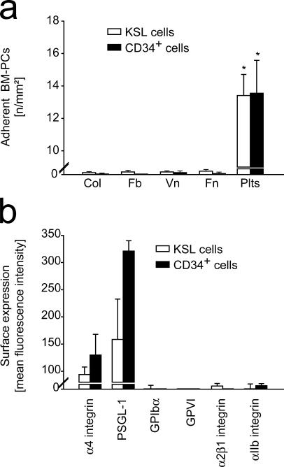
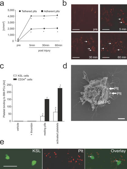
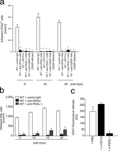
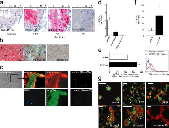
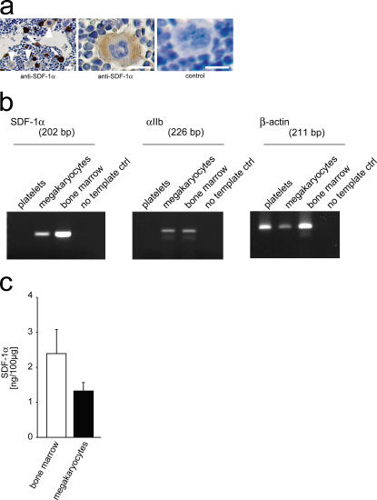
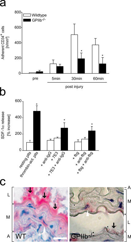
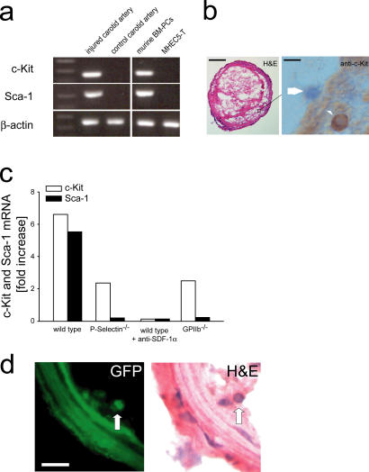
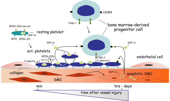
The accumulation of smooth muscle and endothelial cells is essential for remodeling and repair of injured blood vessel walls. Bone marrow-derived progenitor cells have been implicated in vascular repair and remodeling; however, the mechanisms underlying their recruitment to the site of injury remain elusive. Here, using real-time in vivo fluorescence microscopy, we show that platelets provide the critical signal that recruits CD34+ bone marrow cells and c-Kit+ Sca-1+ Lin- bone marrow-derived progenitor cells to sites of vascular injury. Correspondingly, specific inhibition of platelet adhesion virtually abrogated the accumulation of both CD34+ and c-Kit+ Sca-1+ Lin- bone marrow-derived progenitor cells at sites of endothelial disruption. Binding of bone marrow cells to platelets involves both P-selectin and GPIIb integrin on platelets. Unexpectedly, we found that activated platelets secrete the chemokine SDF-1alpha, thereby supporting further primary adhesion and migration of progenitor cells. These findings establish the platelet as a major player in the initiation of vascular remodeling, a process of fundamental importance for vascular repair and pathological remodeling after vascular injury.


Figures

Figure 1.
Figure 1.
Recruitment of BM-PCs to the injured carotid artery. (a) CD34+ BM-PC adhesion before and after carotid injury was monitored by in vivo microscopy. *, P < 0.05 vs. baseline (pre). (b) The microphotographs show representative in vivo fluorescence microscopy images of CD34+ BM-PCs at distinct time points. Bars, 50 μm. (c) Purification of c-Kit+ Sca-1+ Lin PCs. Lin cells were stained with propidium iodide (PI), FITC-conjugated anti–Sca-1 (Ly 6A/E) antibody, and PE-conjugated anti–c-Kit (CD117) antibody. Viable (PI) c-Kit+ Sca-1+ cells were sorted. After sorting, the purity of c-Kit+ Sca-1+ Lin cells was >98%. (d) Assessment of c-Kit+ Sca-1+ Lin BM-PC adhesion before and after carotid injury by in vivo microscopy. *, P < 0.05 vs. no injury. (e) The microphotographs show representative in vivo fluorescence microscopy images of c-Kit+ Sca-1+ Lin BM-PCs before and after vascular injury. Bars represent 50 μm. Arrows in b and e indicate nonadherent BM-PCs, arrowheads indicate adherent BM-PCs. Data are means ± SEM.
Figure 2.
Figure 2.
BM-PCs do not adhere directly to subendothelial matrix proteins under arterial shear conditions. (a) The adhesion of BM-PCs (2 × 10⁴/ml KSL cells or CD34+ cells) to coverslips coated with vitronectin (Vn; Becton Dickinson), collagen (Col; Becton Dickinson), fibrinogen (Fb; Sigma-Aldrich), fibronectin (Fn; Sigma-Aldrich), or to surface adherent platelets was assessed in a transparent flow chamber at a wall shear rate of 1,000 s−1 as described elsewhere (reference 22). The number of adherent BM-PCs is given per mm2 surface. *, P < 0.05 vs. collagen. (b) To determine the adhesion receptors present on the surface of BM-PCs, KSL or CD34+ cells were incubated with fluorophore-labeled anti-αIIb, anti-α4, anti–PSGL-1, anti-α2β1, anti-GPIbα, or anti-GPVI mAb, or irrelevant isotype-matched control antibody and directly analyzed on a FACSCalibur. Data are given as difference between the mean fluorescence intensity [arbitrary units] obtained with the specific antibodies and the signal obtained with the corresponding irrelevant isotype-matched control antibody. Data are means ± SEM.
Figure 3.
Figure 3.
Platelets contribute to the recruitment of BM-PCs to the injured carotid artery. (a) Interactions of rhodamine-6G–tagged platelets before and after carotid injury were investigated by in vivo microscopy. *, P < 0.05 vs. baseline (pre). Representative microscopic images are presented in b. Arrows indicate adherent platelets. (c) BM-PCs were incubated with PBS, thrombin, or resting platelets or platelets activated with thrombin. Flow cytometry was used to detect the binding of CD41+ platelets to BM-PCs. Data are given as increase in mean fluorescence intensity compared with vehicle (PBS)-treated BM-PCs. *, P < 0.05 vs. resting. (d) CD34+ BM-PCs were cultivated in the presence of platelets. Scanning electron microscopy revealed that platelets (arrows) bind directly to the surface of CD34+ BM-PCs. Bar, 5 μm. (e) The carotid artery of C57BL/6J mice was injured, and differentially tagged KSL cells (DCF, green; left) and platelets (rhodamine-6G chloride, red; middle) were visualized by real-time double fluorescence microscopy. The overlay (right) shows that BM-derived c-Kit+ Sca-1+ Lin cells bind exclusively to adherent platelets (arrows). Bars, 50 μm. Data are means ± SEM.
Figure 4.
Figure 4.
Platelet adhesion contributes to the recruitment of BM-PCs to the injured carotid. (a) Carotid injury was induced in wild-type mice treated with function-blocking anti-GPIbα, GPVI-Fc, anti–PSGL-1, anti-CD62P mAbs, or isotype-matched control IgG. In a separate group of animals, carotid injury was induced in mice lacking P-selectin. Adhesion of CD34+ BM-PCs was monitored by in vivo fluorescence microscopy. *, P < 0.05 vs. control IgG-treated WT mice. (b) Carotid injury was induced in wild-type mice treated with function-blocking anti-GPIbα or anti–PSGL-1 mAbs. Adhesion of KSL BM-PCs was monitored by in vivo fluorescence microscopy. *, P < 0.05 vs. control IgG-treated mice. (c) CD34+ BM-PCs were incubated with thrombin-activated platelets in the absence or presence of function-blocking anti–PSGL-1 or anti-CD11b mAb. CD41 expression on the surface of CD34+ BM-PCs was determined to assess platelet binding. Data are means ± SEM.
Figure 5.
Figure 5.
Platelets express and secrete SDF-1α and recruit endogenous PCs to sites of vascular injury. (a) Paraffin-embedded sections of a mouse carotid artery injured as described (panel a and see Materials and methods) and a fresh human coronary thrombus (b) were stained for SDF-1α. Sections incubated with irrelevant isotype-matched IgG served as controls. The intense red (a) or brown (b) staining indicates that SDF-1α is expressed in both mouse and human intravascular thrombi. In a, L, EC, M, and A indicate lumen, endothelial cells, media, and adventitia, respectively. Arrows in b indicate mononuclear cells. Bars, 50 (a) or 20 μm (b). (c) Triple immunofluorescence staining of an injured carotid artery shows that SDF-1α expression in thrombi is largely confined to CD41-expressing platelets. Mononuclear cells recruited to the thrombus are indicated by blue (DAPI). Bars, 25 μm. Sections incubated with the secondary but not the primary antibody served as controls (right). (d) We used ELISA to determine SDF-1α in mouse platelets or whole blood. The mouse melanoma cell line B16-D5 served as control. SDF-1α protein expression is given in nanograms per 100 μg total protein. *, P < 0.05 vs. whole blood. (e) Thrombin-activated human platelets surface express SDF-1α, as indicated by flow cytometry (for details, see Materials and methods). The diagram (left) summarizes three independent experiments and a representative histogram is shown (right). (f) ELISA also demonstrates that platelet activation triggers release of SDF-1α, as indicated by an increase in SDF-1α protein concentration in the supernatants of α-thrombin–activated platelets. *, P < 0.05 vs. resting. (g) Confocal laser scanning microscopy demonstrates that SDF-1α is present in the cytosol of resting platelets, whereas it becomes surface expressed after platelet activation. (top left) Resting platelets at high magnification. SDF-1α is present in the platelet cytoplasm as identified by the red staining (arrowheads); CD41 expression is indicated by green. Platelet activation by 20 μM ADP (top, middle and right) or 0.2 U/ml thrombin (bottom) induced surface mobilization of SDF-1α (green; arrowheads). The platelet cytoskeleton is highlighted by the red staining (phalloidin). Thrombin-activated platelets (bottom right) were stained with irrelevant control mAb (green) and counter-stained with phalloidin (red). Bars, 10 μm (top, middle; bottom, left and right) or 5 μm (top, left and right; bottom, middle). Data are means ± SEM.
Figure 6.
Figure 6.
Megakaryocytes express SDF-1α. (a) Paraffin-embedded sections of mouse femura were stained with anti–SDF-1α. SDF-1α was expressed not only in stromal cells (arrows) but also in megakaryocytes (arrowheads). Bars, 25 μm. (b) Megakaryocytic and platelet SDF-1α mRNA expression was determined using RT-PCR. The mRNA expression of GPIIb integrin and β-actin served as controls. The primer sequences and their corresponding product sizes and annealing temperatures are in Table S1. (c) ELISA was used to determine SDF-1α in mouse megakaryocytes or mouse BM. SDF-1α protein expression is given in nanograms per 100 μg total protein. Data are means ± SEM.
Figure 7.
Figure 7.
GPIIb-dependent platelet aggregation promotes BM-PC recruitment during arterial thrombosis in vivo and contributes to SDF-1α release in vitro. (a) To find out whether platelet GPIIb contributes to BM-PC recruitment in vivo, carotid injury was induced in mice lacking GPIIb. Adhesion of CD34+ BM-PCs was monitored by in vivo fluorescence microscopy. *, P < 0.05 vs. wild type. (b) Platelets were incubated with medium, thrombin, 100 μg/ml soluble fibrinogen, 5 μg/ml mAb anti-fibrinogen, or a combination of both for 30 min. Platelet SDF-1α release was determined by ELISA as described in Materials and methods. Similar experiments were performed with anti-GPIIb mAb 7E3 (whole IgG, 5 μg/ml) in the presence or absence of 5 μg/ml mAb goat anti–mouse IgG. *, P < 0.05 vs. resting platelets. (c) The carotid artery of wild-type (WT) or GPIIb-deficient mice was injured as described in Materials and methods. Paraffin-embedded sections were stained for SDF-1α. L, M, and A indicate lumen, media, and adventitia, respectively. Arrows identify SDF-1α deposited at the site of vascular injury. Bars, 20 μm. Data are means ± SEM.
Figure 8.
Figure 8.
Platelets recruit endogenous PCs to sites of vascular injury. (a) Carotid arteries of C57BL/6J mice were injured as described in Materials and methods. 24 h thereafter, the injured (right) and the uninjured (left, control) carotid arteries were excised and Sca-1 and c-Kit mRNA expression were determined as described in Materials and methods. Isolated mouse CD34+ BM-PCs and the mouse heart EC line MHEC5-T served as positive and negative controls, respectively. The constitutively expressed β-actin transcript was amplified as an internal control to compare relative abundance of PCR products. One representative PCR gel (out of three) is presented. (b) A mouse carotid artery 24 h after vascular injury. Immunohistochemistry demonstrates that c-Kit+ cells are recruited to the luminal aspect of arterial thrombi in vivo (arrowhead). The arrow shows a c-Kit negative cell. Bars, 50 μm (left) and 15 μm (right). (c) Injured carotid arteries of P-selectin–, GPIIb-deficient mice or in anti–SDF-1α mAb-treated wild-type mice were analyzed for c-Kit and Sca-1 mRNA expression. The relative expression of each mRNA was normalized to the expression of β-actin for semiquantification and is presented as fold-increase (mean values) of the injured carotid artery compared with the uninjured control. (d) GFP+ KSL BM-PCs were isolated from the BM of GFP transgenic mice and injected into GFP recipient animals before mechanical injury of the carotid artery. 5 d later, the injured carotid arteries were excised and GFP fluorescence was assessed on cryostat sections. The corresponding hematoxylin and eosin stain of the identical section is shown on the right. Arrows identify GFP+ cells within the neointima. Bars, 25 μm.
Figure 9.
Figure 9.
Adherent platelets recruit BM-PCs to injured vessel wall. Within minutes after vessel injury, platelets adhere to the exposed subendothelium in a process involving GPVI and GPIbα-IX. Adherent/activated platelets surface express P-selectin and release SDF-1α after engagement of GPIIb integrin, thereby initiating BM-PC recruitment within minutes after vessel injury. Within the ensuing hours and days after endothelial disruption, apoptotic SMCs appear to account for the long-term SDF-1α release (reference 25). Hence, SDF-1α delivered by platelets early on and by SMCs at later stages act in concert to promote the entry of BM-PCs at sites of vascular damage.

Similar articles

Cited by

References

    1. Carmeliet, P. 2000. Mechanisms of angiogenesis and arteriogenesis. Nat. Med. 6:389–395. - PubMed
    1. Schwartz, S.M. 1997. Smooth muscle migration in atherosclerosis and restenosis. J. Clin. Invest. 100:S87–S89. - PubMed
    1. Carmeliet, P., L. Moons, J.M. Stassen, M. De Mol, A. Bouche, J.J. van den Oord, M. Kockx, and D. Collen. 1997. Vascular wound healing and neointima formation induced by perivascular electric injury in mice. Am. J. Pathol. 150:761–776. - PMC - PubMed
    1. Werner, N., J. Priller, U. Laufs, M. Endres, M. Bohm, U. Dirnagl, and G. Nickenig. 2002. Bone marrow-derived progenitor cells modulate vascular reendothelialization and neointimal formation: effect of 3-hydroxy-3-methylglutaryl coenzyme a reductase inhibition. Arterioscler. Thromb. Vasc. Biol. 22:1567–1572. - PubMed
    1. Jiang, S., L. Walker, M. Afentoulis, D.A. Anderson, L. Jauron-Mills, C.L. Corless, and W.H. Fleming. 2004. Transplanted human bone marrow contributes to vascular endothelium. Proc. Natl. Acad. Sci. USA. 101:16891–16896. - PMC - PubMed

Publication types

MeSH terms