. 2006 Apr 15;20(8):1028-42.
doi: 10.1101/gad.1406006.

Cross-regulation between Notch and p63 in keratinocyte commitment to differentiation

Affiliations

Cross-regulation between Notch and p63 in keratinocyte commitment to differentiation

Bach-Cuc Nguyen et al. Genes Dev. .

Abstract

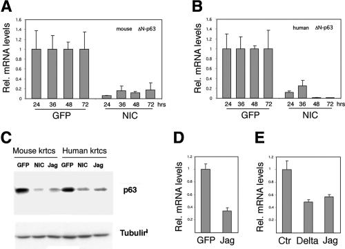
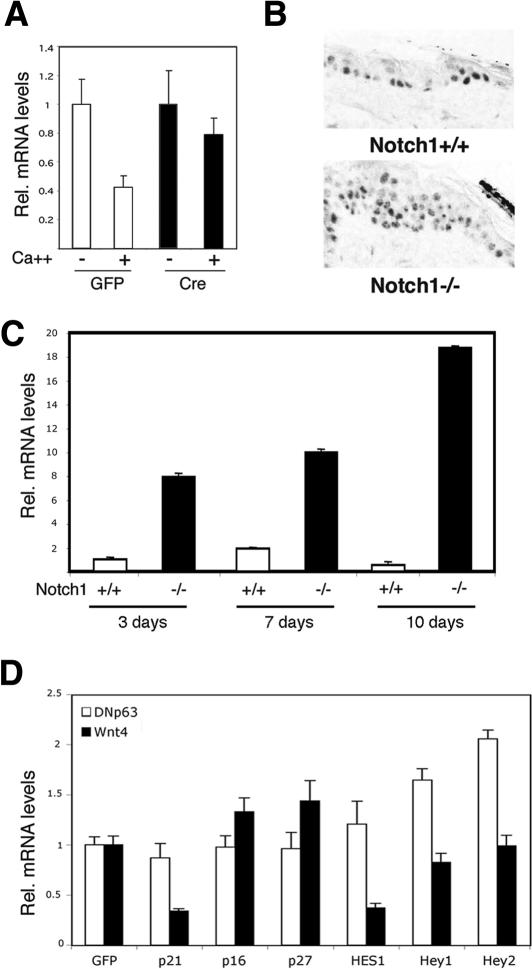
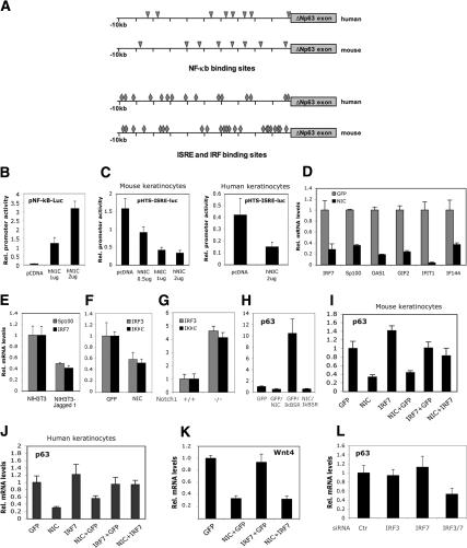
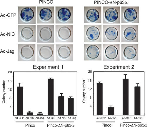
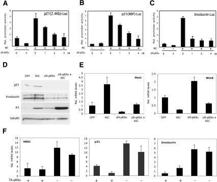
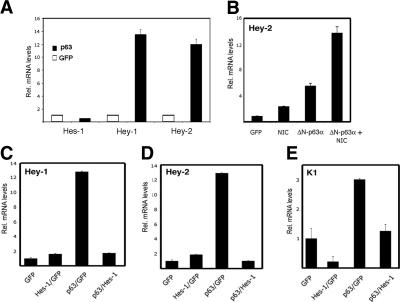
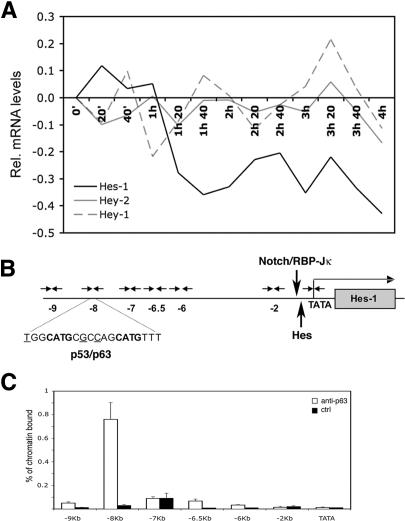
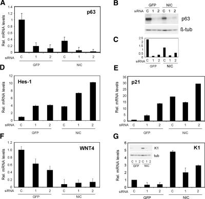
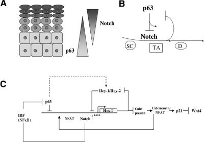
Notch signaling promotes commitment of keratinocytes to differentiation and suppresses tumorigenesis. p63, a p53 family member, has been implicated in establishment of the keratinocyte cell fate and/or maintenance of epithelial self-renewal. Here we show that p63 expression is suppressed by Notch1 activation in both mouse and human keratinocytes through a mechanism independent of cell cycle withdrawal and requiring down-modulation of selected interferon-responsive genes, including IRF7 and/or IRF3. In turn, elevated p63 expression counteracts the ability of Notch1 to restrict growth and promote differentiation. p63 functions as a selective modulator of Notch1-dependent transcription and function, with the Hes-1 gene as one of its direct negative targets. Thus, a complex cross-talk between Notch and p63 is involved in the balance between keratinocyte self-renewal and differentiation.


Figures

Figure 1.
Figure 1.
Negative control of ΔN-p63 expression by increased Notch signaling. (A,B) Down-modulation of ΔN-p63 mRNA expression by activated Notch1. Primary mouse (A) and human (B) keratinocytes were infected with a recombinant adenovirus expressing the cytoplasmic-activated form of Notch1 (NIC) or a control GFP-expressing adenovirus (GFP) for the indicated times (in hours). p63 mRNA levels were quantified by real-time RT–PCR. Values are expressed as relative arbitrary units, after internal normalization for GAPDH (mouse keratinocytes) or β-actin (human keratinocytes) mRNA expression. (C) Down-modulation of ΔN-p63 protein expression by activated Notch1. Primary mouse and human keratinocytes infected with the Ad-GFP (GFP), Ad-NIC (NIC), and Ad Jagged-1 (Jag) adenoviruses were analyzed for levels of p63 protein by immunoblotting with the corresponding antibodies. Immunoblotting for tubulin was used for equal loading control. (D) Down-modulation of p63 mRNA expression in response to increased Jagged 1 expression. Mouse keratinocytes were infected with an adenovirus expressing Jagged 1 (Jag) versus GFP control (GFP) for 24 h followed by p63 mRNA quantification as in the previous panels. (E) Down-modulation of p63 mRNA expression by activation of endogenous Notch in response to Delta 1 or Jagged 1 exposure. Human keratinocytes were cocultured with control mouse NIH3T3 fibroblasts or fibroblasts expressing full-length Delta 1 or Jagged 1 for 48 h, followed by p63 mRNA quantification by RT–PCR with human-specific oligonucleotide primers.
Figure 2.
Figure 2.
Negative control of ΔN-p63 expression in differentiation as a function of endogenous Notch1, separately from cell cycle withdrawal and from p21WAF1/Cip1 and/or Hes-1 expression. (A) Differential down-modulation of ΔN-p63 expression upon induction of differentiation of wild-type versus Notch1−/− keratinocytes. Primary keratinocytes derived from mice with the Notch1 gene flanked by loxP sites (Rangarajan et al. 2001) were infected with a Cre recombinase-expressing adenovirus (Cre) (black bars), for deletion of the Notch1 gene, or with the Ad GFP control (GFP) (white bars). Three days after infection, cells were induced to differentiate by exposure to elevated extracellular calcium for 3 d. p63 mRNA levels were quantified by real-time RT–PCR as in Figure 1. (B) Increased suprabasal p63 expression in the epidermis of mice with an induced deletion of the Notch1 gene. Mice with the Notch1 gene flanked by loxP sites and carrying a keratinocyte-specific K5-CrePR1 transgene versus control Cre-negative littermates were subjected to repeated topical treatments with RU486 for Cre activation as for our previous studies (Mammucari et al. 2005), starting at 5 d of age for five consecutive days. Dorsal skin sections from three mice per group, at 21 d of age, were analyzed by immunohistochemistry with antibodies against p63. Images are representative of a minimum of four independent fields per sections. (C) Up-regulation of ΔN-p63 mRNA expression in the epidermis of mice at early times of Notch1 deletion, prior to any detectable histological alterations. Mice homozygous for the Notch1/loxP gene and carrying the K14CreΔneo transgene (Huelsken et al. 2001) (black bars) versus K14CreΔneo negative controls (white bars) were sacrificed at the indicated days after birth. The epidermis was separated from the underlying dermis by a brief heat shock (30 sec at 60°C) and used for total RNA preparation. p63 mRNA quantification by real-time RT–PCR and GAPDH normalization were carried out as before. Parallel histological analysis of the same skin samples revealed no alterations caused by the Notch1 deletion at 3 and 7 d after birth, with mild hyperplasia becoming detectable at 10 d. The (Notch1/loxP–K14CreΔneo) mice develop substantial skin alterations at later times, similar to those exhibited by mice with an inducible Notch1 deletion (Rangarajan et al. 2001; our unpublished observations). (D) Persistent p63 levels and down-modulation of Wnt4, in keratinocytes with increased p21WAF1/Cip1 and/or Hes-1 expression. Mouse primary keratinocytes were infected with recombinant adenoviruses expressing p21WAF1/Cip1, p27Kip1, p16Ink4a, Hes-1, Hey-1, Hey-2, or GFP control (multiplicity of infection: 100), followed by determination of p63 and Wnt4 mRNA levels by real-time RT–PCR with the corresponding specific primers.
Figure 3.
Figure 3.
Interconnection between Notch, NF-κB, and interferon signaling pathways in control of p63 expression. (A) Schematic of the human and mouse ΔN-p63 promoters with positions of the NF-κB (triangles) and ISRE- and IRF-binding sites (diamonds). Ten kilobases of nucleotide sequence from the transcription initiation site were analyzed by MatInspector 7.4 (Genomatix Software) using an optimized matrix similarity and a core similarity of >0.9. (B) Induction of NF-κB transcriptional activity by activated Notch1. Mouse primary keratinocytes were transfected with a NF-κB-responsive reporter (pNF-κB-luc) with or without increasing amounts of an expression plasmid for activated Notch1 as indicated. Cells were collected 48 h after transfection, and promoter activity values are expressed as arbitrary units using a Renilla reporter for internal normalization. Each condition was tested in triplicate wells, and the standard deviation is indicated. (C) Suppression of interferon response transcriptional activity by activated Notch1 in mouse and human primary keratinocytes (left and right panels, respectively). Cells were transfected with a reporter plasmid carrying multiple copies of an ISRE (pHTS-ISRE-luc) with or without increasing amounts of an expression plasmid for activated Notch1 as indicated. Cells were collected 48 h after transfection, and promoter activity values were calculated using a Renilla reporter for internal normalization as in B. (D) Down-modulation of endogenous interferon-responsive genes in human keratinocytes by activated Notch1 expression. Cells were infected with adenoviruses expressing activated Notch1 (NIC) or GFP-only control (GFP), followed by determination of mRNA expression levels of the indicated genes by real-time RT–PCR analysis. Values are expressed as relative arbitrary units, after internal normalization for β-actin mRNA expression. (E) Down-modulation of IRF7 and Sp100 gene expression by activation of endogenous Notch receptors. Primary human keratinocytes were cocultured with control or Jagged 1-expressing NIH-3T3 fibroblasts as in Figure 1E. IRF7 and Sp100 mRNA levels were determined by real-time RT–PCR as before. (F) Down-modulation of IRF3 and IKKε expression in mouse primary keratinocytes by activated Notch1 expression. Cells were infected with adenoviruses expressing activated Notch1 (NIC) or GFP-only control (GFP), followed by determination of mRNA expression levels by real-time RT–PCR analysis. Values are expressed as relative arbitrary units, after internal normalization for GAPDH mRNA expression. (G) Up-regulation of IRF3 and IKKε expression in the epidermis of mice with an induced deletion of the Notch1 gene. Same RNA samples obtained from 10-d-old mice homozygous for the Notch1/loxP gene and carrying the K14CreΔneo transgene (−/−) versus K14CreΔneo negative controls (+/+) and analyzed for p63 expression in Figure 2C, were analyzed for IRF3 and IKKε mRNA levels by real-time RT–PCR as in the previous panel. (H) Induction of p63 expression by inhibition of NF-κB, with no counteracting effects on suppression by activated Notch1. Primary mouse keratinocytes were infected with recombinant adenoviruses expressing a stabilized form of IκB-α (IκB-SR), activated Notch1 (NIC), or GFP control (GFP), either alone or in various combinations as indicated. p63 mRNA levels were determined by real-time RT–PCR. (I,J) Specific counteracting effects of IRF7 on suppression of p63 expression by activated Notch1. Primary mouse (I) and human (J) keratinocytes were infected with recombinant adenoviruses expressing IRF7 (IRF7), activated Notch1 (NIC), or GFP control (GFP), either alone or in various combinations as indicated. ΔN-p63 mRNA levels were determined by real-time RT–PCR. (K) The same mouse keratinocyte RNA samples as in J were analyzed for levels of Wnt4 expression by real-time RT–PCR with the corresponding specific primers. (L) Down-modulation of p63 expression by knockdown of IRF7 and IRF3 expression. Human primary keratinocytes were transfected with siRNAs targeting the human IRF7 and IRF3 mRNA sequences, individually and in combination, in parallel with scrambled siRNAs control. Cells were analyzed at 48 h after transfection for levels of p63 expression by real-time RT–PCR. Similar results were obtained in two other independent experiments.
Figure 4.
Figure 4.
Counteracting effects of ΔN-p63α on reduction of human keratinocyte proliferative potential by Notch activation. Early-passage primary human keratinocytes were infected with recombinant retroviruses expressing either a full-length ΔN-p63α cDNA together with GFP (PINCO-ΔN-p63α) or GFP alone (PINCO). Three days after infection, GFP-positive cells were purified by sorting and replated. After 3 d of further cultivation, cells were infected with the Ad-GFP, Ad-NIC, or Ad-Jagged 1 viruses. Cells were trypsinized, counted, and replated on triplicate dishes under sparse conditions 6 h after infection; that is, before expression of adenovirally transduced proteins that could interfere with the attachment capability of cells. After 3 wk of further cultivation, clonogenic growth was evaluated by staining of dishes and counting of macroscopically visible colonies (containing >50 cells). Shown is the quantification of this and a second independent experiment.
Figure 5.
Figure 5.
Counteracting effects of ΔN-p63α on the Notch-responsive p21WAF1/Cip1, involucrin, and Hes-1 genes. (AC) Suppression of Notch-dependent transcription in keratinocytes. Primary mouse keratinocytes were transiently transfected with reporter plasmids containing the 2.4-kb promoter region of the p21 gene (A), a minimal Notch-responsive region of the p21 promoter devoid of p53-binding sites but containing a fully conserved RBP-binding site (Rangarajan et al. 2001) (B), or the involucrin promoter (Rangarajan et al. 2001) (C), plus/minus expression plasmids for activated Notch1 (NIC; 1 μg/well), plus/minus an expression vector for ΔN-p63α in increasing amounts as indicated. In all cases, cells were collected 48 h after transfection, and promoter activity values are expressed as arbitrary units using a Renilla reporter for internal normalization. Each condition was tested in triplicate wells, and the standard deviation is indicated. (D,E) Counteracting effects of p63 on endogenous Notch-responsive genes in cultured keratinocytes. Primary mouse keratinocytes were infected with adenoviruses expressing ΔN-p63α (ΔN-p63α) and activated Notch1 (NIC), either individually or in combination. Ad-GFP (GFP) was used as a control and added to the Ad-ΔN-p63α or Ad-NIC viruses when they were used alone, to ensure that in all cases cells received the same amount of viral particles (total multiplicity of infection: 100). Cells were analyzed by either immunoblotting with antibodies against the indicated proteins (D) or real-time RT–PCR with primers specific for the indicated genes (E). (F) Counteracting effects of p63 on endogenous Notch-responsive genes in the skin in vivo. The epidermis from two “gene-switch” TAp63α transgenic mice, in which TAp63α expression was induced in the epidermis by topical application of RU486 (Koster et al. 2004), and two transgenic-negative littermate controls were obtained by laser capture microdissection, followed by total RNA preparation and a single round of linear amplification. Expression of the indicated genes was assessed by real-time RT–PCR with the corresponding specific primers.
Figure 6.
Figure 6.
p63 as a differential modulator of Hes-1 versus Hey-1, Hey-2, and K1 genes. (A) Concomitant induction of Hey-1 and Hey-2 expression with suppression of Hes-1 by increased p63 expression. Mouse primary keratinocytes were infected with a ΔN-p63α (black bars) or GFP control (white bars) adenovirus. Total RNA was prepared from cells at 30 h after infection, followed by real-time RT–PCR analysis using specific oligonucleotide primers for the indicated genes. Values are expressed as relative arbitrary units, after internal normalization for GAPDH mRNA expression. (B) Superinduction of Hey-2 expression by concomitant p63 and activated Notch1 expression. Primary mouse keratinocytes were infected with adenoviruses expressing ΔN-p63α (ΔN-p63α) and activated Notch1 (NIC), either individually or in combination as in Figure 5E. Ad-GFP (GFP) was used as a control and added to the Ad-ΔN-p63α or Ad-NIC viruses when they were used alone, to ensure that in all cases cells received the same amount of viral particles. Cells were analyzed by real-time RT–PCR for Hey-2 mRNA levels. Similar analysis for Hey-1 expression indicated that this gene, unlike Hey-2, is not superinduced by concomitant p63 and NIC expression (data not shown). (C,D) Down-modulation of Hes-1 is required for Hey-1 and Hey-2 induction by p63. Mouse primary keratinocytes were infected with Hes-1 and ΔN-p63α adenoviruses and Ad-GFP control either alone or in combination as indicated. Hey-1 and Hey-2 expression was determined by real-time RT–PCR analysis as in the previous experiments. (E) Down-modulation of Hes-1 is required for K1 induction by p63. The same cells as in the previous experiment were analyzed for levels of keratin 1 (K1) expression by real-time RT–PCR analysis with the corresponding specific primers as indicated.
Figure 7.
Figure 7.
Hes-1 as a direct p63 target gene. (A) Expression profile of Hes-1 (black solid bar), Hey-1 (gray dotted line), and Hey-2 (gray solid bar) at early times upon induction of p63 activity. Primary mouse keratinocytes were infected with a retrovirus carrying an ER-p63 fusion protein (PINCO ER-p63) or empty vector control (PINCO) and subsequently treated with 20 nM tamoxifen for the indicated times. Total RNA was used for cDNA and fluorescent RNA probe preparation followed by hybridization to oligonucleotide microarrays (Mouse Expression Arrays from Affymetrix 430 A 2.0). Data were analyzed using the dChip program (Li and Wong 2001), and values are expressed as changes in relative mRNA levels in the ER-p63-expressing versus control keratinocytes. (B) Map of the Hes-1 promoter. The predicted p53/p63-binding site is indicated together with its precise nucleotide sequence: Bold nucleotides correspond to the core nucleotide sequence required for p53–p63 binding (Barbieri et al. 2005; Ihrie et al. 2005 and references therein), while underlined nucleotides are possible mismatches. The approximate position of the two Notch/RBP-Jκ (−90 and −75) (Jarriault et al. 1995) and four Hes-binding sites (positions −165, −151, −132, and +16) (Takebayashi et al. 1994) is also indicated, together with that of the oligonucleotide primers used for the ChIP analysis (arrows). (C) Specific binding of endogenous p63 to the Hes-1 promoter. Primary mouse keratinocytes under growing conditions were processed for ChIP with antibodies specific for p63 (white bars) or unrelated anti-ERK1 antibodies as control (black bars), followed by real-time PCR amplification of various regions of the Hes-1 promoter indicated in the schematic above. Unprecipitated chromatin preparations were similarly analyzed and used as “input” control. The amount of precipitated DNA was calculated relative to the total input chromatin, and expressed as the percentage of the total according to the following formula (Frank et al. 2001): % total = 2ΔCt × 5, where ΔCt = Ct(input) − Ct(immunoprecipitation). (Ct) Cycle threshold.
Figure 8.
Figure 8.
Control of Hes-1 and other Notch-responsive genes by endogenous p63. (A–C) Knockdown of endogenous p63 expression by siRNA technology. Primary mouse keratinocytes were transfected with siRNAs targeting two distinct regions of the mouse ΔN-p63α mRNA sequence (1 and 2) or scrambled siRNAs control. Parallel cultures were infected with the Ad-GFP and Ad-NIC viruses at 24 h after siRNA transfection as indicated. Cells were analyzed at 48 h after transfection for levels of p63 expression by real-time RT–PCR (A) or immunoblotting with the corresponding antibodies (B). (C) The immunoblotting results were also quantified by densitometric scanning of the autoradiograph and normalization for β-tubulin levels. (D–G) Up-regulation of Hes-1 and p21 expression and down-regulation of Wnt4 and K1 expression as a consequence of p63 knockdown. Primary mouse keratinocyte cultures treated as above were analyzed for expression levels of the indicated genes by real-time RT–PCR analysis and, for K1, also by immunoblotting (G insert).
Figure 9.
Figure 9.
Dynamic model of Notch–p63 cross-regulation in control of keratinocyte self-renewal versus differentiation. (A) Diagram of the epidermis illustrating the inverse gradient of p63 expression versus Notch activity in the lower versus upper epidermal layers, which is likely to result, at least in part, from their reciprocal negative regulation. (B) Scheme illustrating the dual function of p63 in suppressing Notch signaling in epidermal cells with high self-renewal potential, while synergizing with other specific aspects of Notch function involved in the early stages of differentiation. (SC) Putative stem cell populations; (TA) transient amplifying cells. Down-modulation of p63 expression by increased Notch signaling could then be a signal for later stages to occur. (D) Differentiated cells. (C) The Hes-1 gene as a relay for multiple feedback mechanisms in the integrated control of keratinocyte self-renewal versus differentiation. While Hes-1 is a direct target of p63, with its down-modulation leading to an induction of Hey-1 and Hey-2 family members, we have found that Hey1/2 overexpression, in turn, down-modulates Hes-1 levels, providing a possible reinforcement mechanism for the negative regulation of Hes-1 by p63. The other signaling pathways that converge on control of the Hes-1 gene and its downstream involvement in regulating calcineurin/NFAT activity and p21 and Wnt4 expression are discussed in the text.

References

    1. Artavanis-Tsakonas S., Rand M.D., Lake R.J. Notch signaling: Cell fate control and signal integration in development. Science. 1999;284:770–776. - PubMed
    1. Barbieri C.E., Perez C.A., Johnson K.N., Ely K.A., Billheimer D., Pietenpol J.A. IGFBP-3 is a direct target of transcriptional regulation by ΔNp63α in squamous epithelium. Cancer Res. 2005;65:2314–2320. - PubMed
    1. Barrandon Y., Green H. Three clonal types of keratinocyte with different capacities for multiplication. Proc. Natl. Acad. Sci. 1987;84:2302–2306. - PMC - PubMed
    1. Bernassola F., Oberst A., Melino G., Pandolfi P.P. The promyelocytic leukaemia protein tumour suppressor functions as a transcriptional regulator of p63. Oncogene. 2005;24:6982–6986. - PubMed
    1. Capobianco A.J., Zagouras P., Blaumueller C.M., Artavanis-Tsakonas S., Bishop J.M. Neoplastic transformation by truncated alleles of human NOTCH1/TAN1 and NOTCH2. Mol. Cell. Biol. 1997;17:6265–6273. - PMC - PubMed

Publication types