. 2006 Apr 24;173(2):241-51.
doi: 10.1083/jcb.200506163. Epub 2006 Apr 17.

Synaptotagmin IV is necessary for the maturation of secretory granules in PC12 cells

Affiliations

Synaptotagmin IV is necessary for the maturation of secretory granules in PC12 cells

Malika Ahras et al. J Cell Biol. .

Abstract

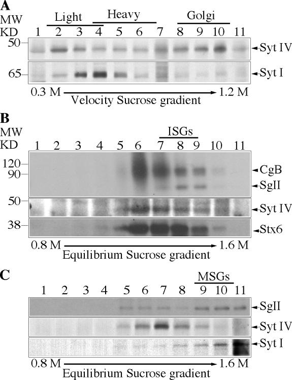
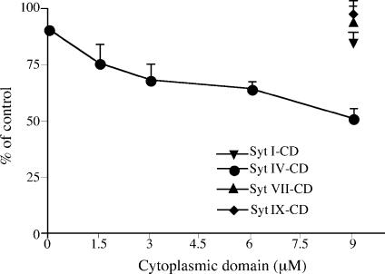
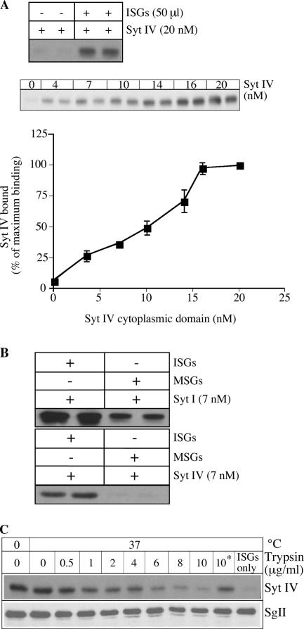
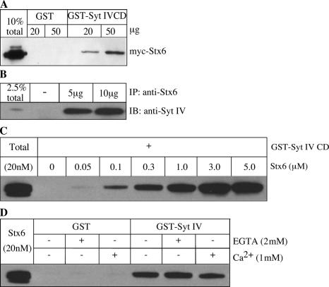
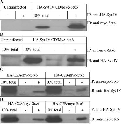
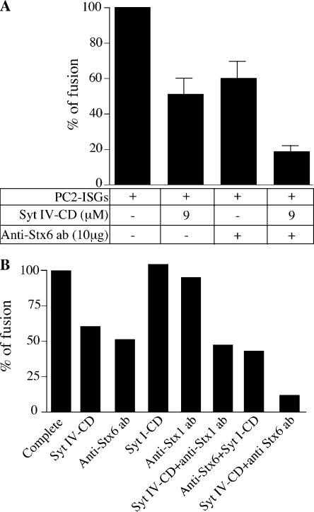
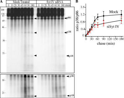
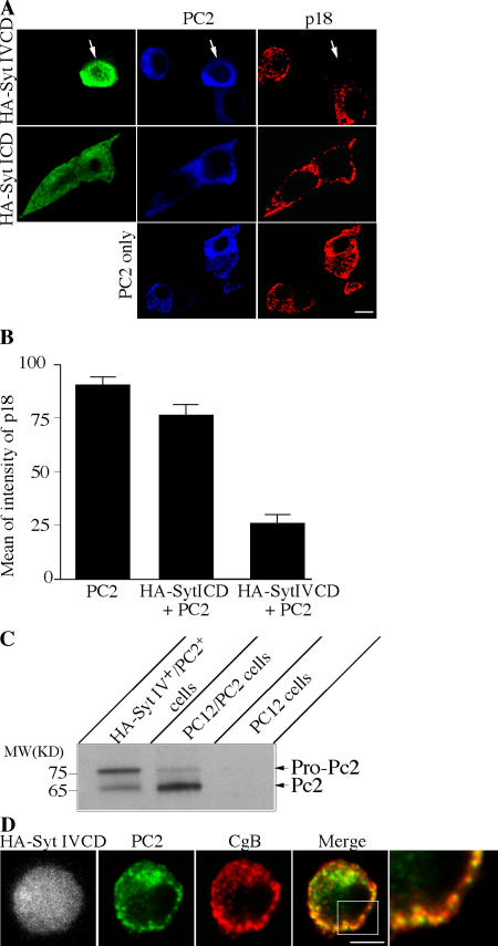
In neuroendocrine PC12 cells, immature secretory granules (ISGs) mature through homotypic fusion and membrane remodeling. We present evidence that the ISG-localized synaptotagmin IV (Syt IV) is involved in ISG maturation. Using an in vitro homotypic fusion assay, we show that the cytoplasmic domain (CD) of Syt IV, but not of Syt I, VII, or IX, inhibits ISG homotypic fusion. Moreover, Syt IV CD binds specifically to ISGs and not to mature secretory granules (MSGs), and Syt IV binds to syntaxin 6, a SNARE protein that is involved in ISG maturation. ISG homotypic fusion was inhibited in vivo by small interfering RNA-mediated depletion of Syt IV. Furthermore, the Syt IV CD, as well as Syt IV depletion, reduces secretogranin II (SgII) processing by prohormone convertase 2 (PC2). PC2 is found mostly in the proform, suggesting that activation of PC2 is also inhibited. Granule formation, and the sorting of SgII and PC2 from the trans-Golgi network into ISGs and MSGs, however, is not affected. We conclude that Syt IV is an essential component for secretory granule maturation.


Figures

Figure 1.
Figure 1.
Syt IV is found on the Golgi and ISGs in PC12 cells. (A) Fractions from a 0.3–1.2 M continuous velocity sucrose gradient loaded with a PNS from PC12 cells were collected and analyzed with anti–Syt IV (top) and Syt I (bottom) antibodies. Alternatively, PC12 cells were labeled with [35S]sulfate for 5 min and chased for 15 min (B) or labeled for 1 h and chased O/N (C) to label ISGs or MSGs, respectively, and then subjected to velocity centrifugation. (B and C) Fractions 1–4 (B) or 4–6 (C) from the velocity gradients were further separated on a 0.8–1.6 M discontinuous equilibrium sucrose gradient and analyzed. (B and C, top) Autoradiogram showing [35S]sulfate-labeled CgB (B) and SgII (B and C). (B and C, bottom) Western blot analysis using anti–Syt IV and anti-Stx6 antibodies (B) or anti–Syt IV and anti–Syt I antibodies (C). In B, the diffuse band between 120 and 90 kD is a heparan sulfate proteoglycan (Tooze and Huttner, 1990).
Figure 2.
Figure 2.
The CDs of Syt IV, but not of Syt I, VII, or IX, inhibit ISG homotypic fusion. An ISG homotypic fusion assay was performed as previously described by Urbé et al. (1998), which measures fusion by the production of p18 from [35S]sulfate-labeled SgII. The complete fusion reaction contains PC2 ISGs, PC12 [35S]sulfate-labeled PNS, and ATP. Syt IV (circle), Syt I (inverted triangle), Syt VII (triangle), or Syt IX (diamond) CDs were added at the indicated concentrations, on ice, and then the fusion assay was performed. After subtraction of the background from PC2-ISG minus reactions, the fusion was assayed by quantifying the amount of p18 produced and is presented as a percentage of the complete fusion reaction. The data represent the mean of three experiments done in duplicate. Error bars are the SEM. The addition of more Syt IV CD did not increase the inhibition beyond 50% (not depicted).
Figure 3.
Figure 3.
Syt IV CD is recruited to ISG membranes. (A) 20 nM of Syt IV CD was incubated for 30 min at 37°C with or without 50 μl PC12 ISGs, or increasing amounts of Syt IV CD were added to 50 μl ISGs in fusion buffer with ATP. The amount of Syt IV bound to ISG membranes was determined by immunoblotting with anti–Syt IV antibody and quantified using ImageJ software. The data are the mean of three experiments done in duplicate. Error bars are the SEM. (B) 7 nM Syt IV or Syt I CDs were preincubated with 50 μl ISGs or MSGs in fusion buffer with ATP. The amount of Syt IV or Syt I bound was determined as described in Materials and methods. (C) 50 μl ISGs were preincubated with increasing amounts of trypsin. Trypsin inhibitor was added before (*) or after the preincubation. Binding reactions including 18 nM Syt IV CD were performed as described in Materials and methods. The amount of Syt IV bound to ISGs was detected with an anti–Syt IV antibody (top). The blot was stripped and reprobed with an anti-SgII antibody (bottom).
Figure 4.
Figure 4.
Syt IV interacts with Stx6. (A) Cell lysates from HEK cells transfected with myc-Stx6 were incubated with GST-Syt IV CD or GST bound to glutathione beads. (B) PC12 cells were lysed in TNTE buffer and incubated with 5 or 10 μg of anti-Stx6 antibody overnight at 4°C, and subsequently bound to protein G beads. Immunoprecipitates and 2.5% of the input were analyzed by immunoblotting with anti–Syt IV antibody. (C) Direct binding of Stx6 to Syt IV in vitro. 3 μg of GST-Syt IV CD bound to glutathione beads was incubated with the indicated amounts of the Stx6 CD in binding buffer overnight, and bound protein was analyzed by immunoblotting with anti-Stx6 antibody 3D10. (D) The Syt IV–Stx6 interaction in vitro is independent of Ca2+. Binding assays were performed as in C, using 1 μM Stx6 CD, supplemented with either 1 mM CaCl2 or 2 mM EGTA.
Figure 5.
Figure 5.
Syt IV interacts with Stx6 via both C2A and C2B domains. (A and B) HEK293 cells were cotransfected with HA-Syt IV CD and myc-Stx6. Lysates from untransfected and transfected cells were used for immunoprecipitation with anti-HA (+) or beads alone (−; A) and with anti-myc antibody or beads alone (B). Immunoprecipitates and 10% of total input were analyzed with anti-myc (A) or anti-HA (B) antibodies by immunoblotting. (C and D) Lysates from HEK293 cells cotransfected with either HA-Syt IV C2A or C2B domain and myc-Stx6 were subjected to immunoprecipitation with either anti-myc antibody or beads alone (C), blotted with anti-HA antibody or anti-HA antibody or beads alone (D), and then blotted with anti-myc antibody.
Figure 6.
Figure 6.
Syt IV CD and Stx6 antibody have an additive inhibitory effect on ISG homotypic fusion. (A) A complete fusion reaction was incubated with 9 μM Syt IV CD and 10 μg anti-Stx6 antibody, or Syt IV CD and anti-Stx6 antibody. Fusion reactions were performed, treated, and analyzed (see Materials and methods). p18 signals were quantified using ImageJ analysis software, and the background signal measured in the absence of PC2 ISGs was subtracted. The data is the mean of three independent experiments done in duplicate. Error bars are the SEM. (B) A complete fusion reaction was incubated with 9 μM Syt IV CD, 10 μg anti-Stx6 antibody, 9 μM Syt I CD, 10 μg anti-Stx1 antibody, Syt IV CD and anti-Stx1 antibody, 9 μM Syt I CD and 10 μg anti-Stx6 antibody, or 10 μg anti-Stx6 antibody and 9 μM Syt IV CD. p18 signals were quantified as described in A.
Figure 7.
Figure 7.
siRNA-mediated depletion of Syt IV results in inhibition of ISG fusion. PC12/PC2 cells were either transfected with 50 nM Syt IV siRNA or mock-transfected with transfection reagents alone. (A) 72 h after transfection, cells were labeled with anti–Syt IV (green) or anti–Syt I (blue). Images were taken using a Zeiss LSM510 confocal microscope. Bars, 20 μm. (B) 10 μg mock- or siRNA-treated cell lysates were solubilized and analyzed with anti–Syt IV and anti–Syt I antibodies. (C) Scheme for the pulse-chase labeling protocol and analysis of ISG size used in D and E, as well as in Fig. 8 A (Tooze et al., 1991). (D) 72 h after mock or Syt IV siRNA treatment, PC12 cells were labeled, harvested, and analyzed by velocity-controlled sucrose gradient fractionation. Fractions 1–12 were analyzed from gradients loaded with PNS prepared from mock- or siRNA-treated cells after a 60-min chase. In mock-treated cells, SgII is found in ISGs sedimenting in fractions 2–6, whereas in Syt IV siRNA-treated cells ISGs containing SgII are found in fractions 1–4. (E) Quantitation of the position of SgII in fractions 1–6 from velocity gradients loaded with cells treated as in D and chased for 30 and 60 min. The average of two experiments is shown.
Figure 8.
Figure 8.
Syt IV depletion inhibits SgII processing in PC12/PC2 cells. (A) Syt IV siRNA– or mock-transfected PC12/PC2 cells were pulse-labeled with [35S]sulfate for 5 min and chased for the indicated length of time. Cells were lysed in TNTE and a heat-stable fraction was prepared and analyzed by SDS-PAGE gel and autoradiography. Arrowheads indicate the position of the SgII-processing products p38 and p18. (bottom) The same gel presented in the top image, but at higher exposure. (B) Quantification of the ratio of p38 and p86 using ImageJ. Three separate experiments were analyzed and the average is shown. Error bars are the SEM. P < 0.02, paired t test.
Figure 9.
Figure 9.
Syt IV CD inhibits PC2 maturation and PC2-dependent SgII processing. (A) PC12 cells were cotransfected with HA-Syt IV CD and PC2, HA-Syt I CD and PC2, or PC2 alone. The cells were fixed and labeled with rat anti-HA (green), rabbit anti-PC2 (blue), and mouse anti-p18 (red). (B) Transfected cells were individually selected and the mean of intensity of each channel was measured. Columns represent average of the mean of intensity of each channel. Error is the SEM, where n = 22 cells for all conditions. (C) PC12 cells were cotransfected with HA-Syt IV CD and PC2. Cells positive for both HA-Syt IV and PC2 were FACS-sorted by labeling with anti-HA antibody (Alexa Fluor 488) and anti-PC2 antibody (Alexa Fluor 647). As a control, PC12/PC2 cells labeled for PC2 were FACS-sorted using identical conditions. 150,000 HA-Syt IV+/PC2+ cells and 300,000 PC12/PC2 cells were loaded on an SDS-PAGE gel and immunoblotted with the anti-PC2 antibody. 5 μg of a PC12 cell lysate was used as a negative control for the PC2 antibody. (D) PC12 cells cotransfected with HA-Syt IV CD and PC2 were fixed and labeled with anti-HA, anti-PC2, and chromogranin B (CgB) antibodies. Bars, 5 μm.

Similar articles

Cited by

References

    1. Adolfsen, B., S. Saraswati, M. Yoshihara, and J.T. Littleton. 2004. Synaptotagmins are trafficked to distinct subcellular domains including the postsynaptic compartment. J. Cell Biol. 166:249–260. - PMC - PubMed
    1. Arvan, P., and D. Castle. 1998. Sorting and storage during secretory granule biogenesis: looking backward and looking forward. Biochem. J. 332:593–610. - PMC - PubMed
    1. Austin, C., I. Hinners, and S.A. Tooze. 2000. Direct and GTP-dependent interaction of ADP-ribosylation factor 1 with clathrin adaptor protein AP-1 on immature secretory granules. J. Biol. Chem. 275:21862–21869. - PubMed
    1. Bai, J., and E.R. Chapman. 2004. The C2 domains of synaptotagmin–partners in exocytosis. Trends Biochem. Sci. 29:143–151. - PubMed
    1. Bennett, M.K., N. Calakos, and R.H. Scheller. 1992. Syntaxin: a synaptic protein implicated in docking of synaptic vesicles at presynaptic active zones. Science. 257:255–259. - PubMed