. 2006 Apr 25;103(17):6735-40.
doi: 10.1073/pnas.0510645103. Epub 2006 Apr 17.

Activity-dependent release of precursor nerve growth factor, conversion to mature nerve growth factor, and its degradation by a protease cascade

Affiliations

Activity-dependent release of precursor nerve growth factor, conversion to mature nerve growth factor, and its degradation by a protease cascade

Martin A Bruno et al. Proc Natl Acad Sci U S A. .

Abstract

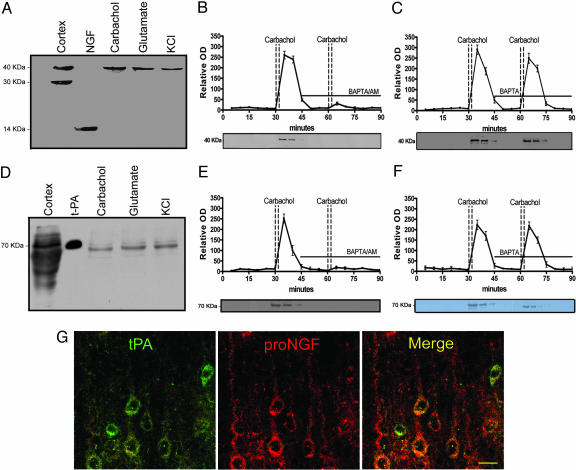
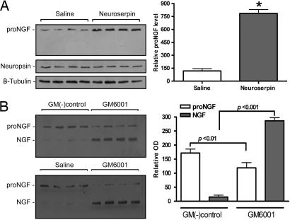
In this report, we provide direct demonstration that the neurotrophin nerve growth factor (NGF) is released in the extracellular space in an activity-dependent manner in its precursor form (proNGF) and that it is in this compartment that its maturation and degradation takes place because of the coordinated release and the action of proenzymes and enzyme regulators. This converting protease cascade and its endogenous regulators (including tissue plasminogen activator, plasminogen, neuroserpin, precursor matrix metalloproteinase 9, and tissue inhibitor metalloproteinase 1) are colocalized in neurons of the cerebral cortex and released upon neuronal stimulation. We also provide evidence that this mechanism operates in in vivo conditions, as the CNS application of inhibitors of converting and degrading enzymes lead to dramatic alterations in the tissue levels of either precursor NGF or mature NGF. Pathological alterations of this cascade in the CNS might cause or contribute to a lack of proper neuronal trophic support in conditions such as cerebral ischemia, seizure and Alzheimer's disease or, conversely, to excessive local production of neurotrophins as reported in inflammatory arthritis pain.


Conflict of interest statement

Conflict of interest statement: No conflicts declared.

Figures

Fig. 1.
Fig. 1.
Neuronal colocalization and stimulus-coupled release of proNGF and tPA. Western blots demonstrating proNGF (A) and tPA (D) released from cerebral cortex after stimulation (see Materials and Methods). First lane illustrates immunoreactive bands from cortical homogenates. In the second lane, 5 ng mNGF (A) or 3 ng tPA (D) are loaded as control. Time course of proNGF (B) and tPA (E) released from cerebral cortex tissue. Activity-dependent release of neuroactive proteins was induced by two consecutive carbachol stimulations. The presence of the intracellular calcium chelator, BAPTA/AM (10 μM), in the superfusion buffer inhibited the release of proNGF (B) and of tPA (E) but the presence of the extracellular calcium chelator, BAPTA (10 μM), did not affect proNGF (C) or tPA (F) release (mean ± SEM). (G) Localization of tPA (green) and proNGF (red) in cortical pyramidal neurons; colocalization illustrated with merged images (yellow). (Scale bar: 20 μm.)
Fig. 2.
Fig. 2.
Plasminogen and neuroserpin release conditions. Western blots of plasminogen (A) and neuroserpin (B) released after carbachol, glutamate, and KCl stimulation. The first lane illustrates immunoreactive bands from cortical homogenates; the second lane shows positive controls for mouse plasminogen (10 ng) and neuroserpin (6 ng), respectively.
Fig. 3.
Fig. 3.
Colocalization of tPA with plasminogen, neuroserpin, proMMP-9, and TIMP-1 at pyramidal neurons. (Left) tPA immunolocalization is shown in green. (Center) Immunolocalization of plasminogen, neuroserpin, proMMP9, and TIP-1 are shown in red. (Right) Merged images with tPA on left column; yellow indicates co-localization in the same neuron. (Scale bar: 20 μm.)
Fig. 4.
Fig. 4.
Activity-dependant release of proMMP-9 and TIMP-1. Representative Western blots of proMMP-9 (A) and TIMP-1 (B) released by cerebral cortical tissue upon stimulation with carbachol (100 nM), glutamate (60 μM), or KCl (50 mM).
Fig. 5.
Fig. 5.
Maturation of released proNGF and degradation of mature NGF. (A) Plasmin-induced conversion of endogenously released proNGF into mNGF. The plasmin protease activity on proNGF was inhibited by α2-antiplasmin. MMP-2, MMP-7, and MMP-9 failed to convert proNGF into mNGF. (B) tPA, plasminogen, or neuroserpin alone (lanes 3, 4 and 7, respectively) were not able to convert proNGF on their own; it was only when both plasminogen and tPA were present that the conversion took place (lane 5). The convertase activity was inhibited by neuroserpin (lane 6). NGF (A, B, and D, lane 1) and proNGF (A and B, lane 2) are reference controls (C). MMP-9 alone was not able to degrade mNGF (B, lane 2) whereas NGF degradation occurred in the presence of either MMP-9, tPA, and plasminogen (lane 5) or with MMP-9 plus plasmin (lane 6). (D) Neither plasmin nor proMMP-9 alone degraded mNGF (lane 2 and 3, respectively). The plasmin activated MMP-9 degradation of mNGF (lane 4) was blocked by the metalloproteinase inhibitor GM6001 (lane 5) but not by the negative control GM6001 (lane 6).
Fig. 6.
Fig. 6.
The cortical proNGF/matureNGF ratio is changed by the application of neuroserpin or MMP-9 inhibitors. (A) Increased amount of cortical proNGF in neuroserpin-treated animals (mean ± SEM; P < 0.001; t test). (B) The inhibitor of matrix metalloproteinase GM6001 significantly increased the cortical mNGF (P < 0.001) and decreased proNGF (P < 0.01) when compared with the GM6001 negative or saline control-treated (mean ± SEM). The levels of neuropsin, a serine protease secretory protein present in pyramidal neurons and β-tubulin were not altered in these experiments.
Fig. 7.
Fig. 7.
Schematic representations of events leading to proNGF conversion into mNGF and its degradation. Neuronally stored proNGF, plasminogen, tPA, neuroserpin, proMMP-9, and TIMP-1 would be released into the extracellular space upon neuronal stimulation. Released tPA would induce the conversion of plasminogen to plasmin, where its activity is tightly regulated by secreted neuroserpin. The generated plasmin would convert proNGF into mature NGF and activate proMMP-9 into active MMP-9. Mature NGF would interact with its cognate receptors (TrkA and p75 neurotrophin receptor) or suffer degradation by activated MMP-9.

Similar articles

Cited by

References

    1. Bibel M., Barde Y. A. Genes Dev. 2000;14:2919–2937. - PubMed
    1. Levi-Montalcini R. Science. 1987;237:1154–1162. - PubMed
    1. Thoenen H. Science. 1995;270:593–598. - PubMed
    1. Sofroniew M. V., Galletly N. P., Isacson O., Svendsen C. N. Science. 1990;247:338–342. - PubMed
    1. Debeir T., Saragovi H. U., Cuello A. C. Proc. Natl. Acad. Sci. USA. 1999;96:4067–4072. - PMC - PubMed

Publication types

LinkOut - more resources