Comparative Study
. 2006 Apr 25;103(17):6471-6.
doi: 10.1073/pnas.0511020103. Epub 2006 Apr 17.

Crystal structures of gamma-glutamyltranspeptidase from Escherichia coli, a key enzyme in glutathione metabolism, and its reaction intermediate

Affiliations
Comparative Study

Crystal structures of gamma-glutamyltranspeptidase from Escherichia coli, a key enzyme in glutathione metabolism, and its reaction intermediate

Toshihiro Okada et al. Proc Natl Acad Sci U S A. .

Abstract

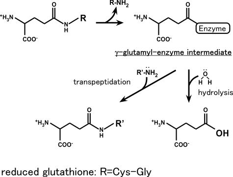
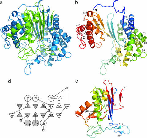
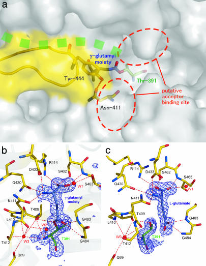
Gamma-glutamyltranspeptidase (GGT) is a heterodimic enzyme that is generated from the precursor protein through posttranslational processing and catalyzes the hydrolysis of gamma-glutamyl bonds in gamma-glutamyl compounds such as glutathione and/or the transfer of the gamma-glutamyl group to other amino acids and peptides. We have determined the crystal structure of GGT from Escherichia coli K-12 at 1.95 A resolution. GGT has a stacked alphabetabetaalpha fold comprising the large and small subunits, similar to the folds seen in members of the N-terminal nucleophile hydrolase superfamily. The active site Thr-391, the N-terminal residue of the small subunit, is located in the groove, from which the pocket for gamma-glutamyl moiety binding follows. We have further determined the structure of the gamma-glutamyl-enzyme intermediate trapped by flash cooling the GGT crystal soaked in glutathione solution and the structure of GGT in complex with l-glutamate. These structures revealed how the gamma-glutamyl moiety and l-glutamate are recognized by the enzyme. A water molecule was seen on the carbonyl carbon of the gamma-glutamyl-Thr-391 Ogamma bond in the intermediate that is to be hydrolyzed. Notably the residues essential for GGT activity (Arg-114, Asp-433, Ser-462, and Ser-463 in E. coli GGT) shown by site-directed mutagenesis of human GGT are all involved in the binding of the gamma-glutamyl moiety. The structure of E. coli GGT presented here, together with sequence alignment of GGTs, may be applicable to interpret the biochemical and genetic data of other GGTs.


Conflict of interest statement

Conflict of interest statement: No conflicts declared.

Figures

Scheme 1.
Scheme 1.
Reactions catalyzed by GGT.
Fig. 1.
Fig. 1.
Structure of E. coli GGT. (a) Ribbon drawing of the GGT heterodimer. The L subunit is colored blue, and the S subunit is colored green. (b) Ribbon drawing of the L subunit. (c) Ribbon drawing of the S subunit. α-Helices and β-strands are labeled. In each of the L and S subunits, the N terminus is blue and the C terminus is red, with intermediate colors following the distance in the sequence from the N terminus. The N-terminal residue of the S subunit (Thr-391) is shown with a stick model. (d) A topology diagram of E. coli GGT. Circle, triangle, and square indicate α-helix, β-strand, and insertion not conserved among Ntn-hydrolases, respectively. The secondary structures were defined with dssp (19). The figures were prepared with pymol (20) and tops (21).
Fig. 2.
Fig. 2.
The structure of the substrate binding pocket of E. coli GGT. (a) Surface drawing of substrate binding pocket. The stick model of the γ-glutamyl moiety, nucleophile (Thr-391), and residues forming the wall (Asn-411 and Tyr-444) are shown in blue, green, and yellow, respectively. Green dots represent the groove in which the peptide of the precursor protein is assumed to be present. The hydrogen bond between Asn-411 Oδ and Tyr-444 Oη is shown as a dashed line. The ribbon model shown in yellow represents residues Pro-438–Gly-449, which are absent in B. subtilis GGT. (b) The (FoFc) omit map contoured at the 3σ level for GGT-γG. The omit map was generated by omitting the γ-glutamyl moiety, Thr-391, and a water molecule (labeled W2) from the model. Ball-and-stick models of γ-glutamyl–enzyme complex are overlaid on the map. The residues involved in substrate binding and enzyme reaction are shown in the model. For the clarity, the side chains of Gln-89, Leu-410, and Thr-412 are omitted from the model. Water molecules involved in substrate binding and the catalytic reaction are labeled (W1∼W3). The hydrogen bonds are shown as dashed lines. (c) The (FoFc) omit map for GGT-Glu prepared as for GGT-γG. The view direction is rotated by 40° around the vertical axis relative to b. The figures were prepared with pymol (20).
Fig. 3.
Fig. 3.
Multiple sequence alignment of GGT of several representative organisms. The sequence numbering corresponds to E. coli GGT. The secondary structure of E. coli GGT is shown above the sequence. Identical residues are shaded, and similar residues are boxed. The residues of catalytic nucleophile, stabilizing the nucleophile, substrate binding, and comprising the oxyanion hole are shown with the letters N, S, B, and O, respectively. The residues that form the substrate-binding pocket wall (Asn-411 and Tyr-444 in E. coli GGT) are shown as triangles, and the segment from Pro-438 to Gly-449 in E. coli is shown in the gray bar. The residues that significantly reduced enzymatic activity by site-directed mutagenesis for human GGT are underlined. Sequences shown are for GGTs of E. coli (ECOLI), B. subtilis (BACSU), human, and rat. The figure was prepared with clustalw (25) and espript (26).

Similar articles

Cited by

References

    1. Tate S. S., Meister A. Mol. Cell. Biochem. 1981;39:357–368. - PubMed
    1. Taniguchi N., Ikeda Y. Adv. Enzymol. Relat. Areas Mol. Biol. 1998;72:239–278. - PubMed
    1. Hanigan M. H., Ricketts W. A. Biochemistry. 1993;32:6302–6306. - PubMed
    1. Suzuki H., Hashimoto W., Kumagai H. J. Bacteriol. 1993;175:6038–6040. - PMC - PubMed
    1. Mehdi K., Penninckx M. J. Microbiology. 1997;143:1885–1889. - PubMed

Publication types

LinkOut - more resources