. 2006 Apr 25;103(17):6730-4.
doi: 10.1073/pnas.0509765103. Epub 2006 Apr 17.

Identification of a structural motif crucial for infectivity of hepatitis B viruses

Affiliations

Identification of a structural motif crucial for infectivity of hepatitis B viruses

Lars Stoeckl et al. Proc Natl Acad Sci U S A. .

Abstract

Infectious entry of hepatitis B viruses (HBV) has nonconventional facets. Here we analyzed whether a cell-permeable peptide [translocation motif (TLM)] identified within the surface protein of human HBV is a general feature of all hepadnaviruses and plays a role in the viral life cycle. Surface proteins of all hepadnaviruses contain conserved functional TLMs. Genetic inactivation of the duck HBV TLMs does not interfere with viral morphogenesis; however, these mutants are noninfectious. TLM mutant viruses bind to cells and are taken up into the endosomal compartment, but they cannot escape from endosomes. Processing of surface protein by endosomal proteases induces their exposure on the virus surface. This unmasking of TLMs mediates translocation of viral particles across the endosomal membrane into the cytosol, a prerequisite for productive infection. The ability of unmasked TLMs to translocate processed HBV particles across cellular membranes was shown by confocal immunofluorescence microscopy and by infection of nonpermissive cell lines with HBV processed in vitro with endosomal lysate. Based on these data, we propose an infectious entry mechanism unique for hepadnaviruses that involves virus internalization by receptor-mediated endocytosis followed by processing of surface protein in endosomes. This processing activates the function of TLMs that are essential for viral particle translocation through the endosomal membrane into the cytosol and productive infection.


Conflict of interest statement

Conflict of interest statement: No conflicts declared.

Figures

Fig. 1.
Fig. 1.
Destruction of the TLM abolishes infectivity of DHBV. (A) Immunofluorescence microscopy of infected PDH using an L-specific antiserum. Cells were infected with 100 MGE WT DHBV, DHBVD2, or DHBVD1/2 mutant. Cells were fixed 4 days after infection. Hoechst staining was used to visualize nuclei. The photographs were taken at ×200 magnification. (B) Immunoblot analysis of lysates from PDHs infected with WT DHBV or the mutants by using a DHBV core-specific antiserum. Uninfected PDHs served as negative control. (C) PDHs were infected with 100 MGE. Cells were harvested 7 days after infection and analyzed for replicative intermediates by Southern blotting. (D) Analysis of cccDNA by PCR. The cccDNA was isolated 3 days after infection and amplified by PCR by using cccDNA-selective primers. Uninfected PDHs served as negative control.
Fig. 2.
Fig. 2.
TLM-deficient viral particles are trapped in the endosome. PDH were inoculated with WT or mutant DHBV (200 MGE). After 10 h of incubation with WT DHBV, DHBVD1/2, DHBVD2, and DHBVD1, cells were harvested and subfractionated. Cytosolic and endosomal fractions were adjusted to identical protein concentrations, and their purity was controlled by immunoblotting by using grb2 (cytoplasm)- and clathrin HC (endosomes)-specific antisera. For detection of viral DNA in the cytosolic (c) and endosomal (e) fractions, TaqMan PCR was performed. The y axis indicates the number of viral genomes per 25 μl of resuspended subcellular fraction.
Fig. 3.
Fig. 3.
Cleavage of HBsAg by endosomal proteases results in surface exposure of the TLM. Purified HBV particles were subjected to immunoprecipitation with either an HBV-TLM-specific antiserum (lanes 2, 4, 6, and 8), or an HBsAg-specific serum as a positive control (lanes 1, 3, 5, and 7). The precipitated material was immunoblotted by using an HBcAg-specific antiserum. In lanes 1 and 2 precipitates of untreated HBV particles were loaded using an HBsAg-specific serum or a TLM-specific serum. Purified HBV particles were incubated for 30 min at pH 5.0 and immunoprecipitated by using an HBV-TLM-specific antiserum (lane 4) and an HBsAg-specific serum (lane 3). Purified HBV particles were incubated for 30 min at pH 5.0, then by addition of 10× PBS the pH was shifted to ≈7. Afterward, immunoprecipitation was performed by using an HBV-TLM-specific antiserum (lane 6) and an HBsAg-specific serum (lane 5). Purified HBV particles were incubated for 30 min at pH 5.0 in endosomal lysate from HepG2 cells and immunoprecipitated by using an HBV-TLM-specific antiserum (lane 8) and an HBsAg-specific serum (lane 7). Recombinant HBcAg (lane 9) served as positive control.
Fig. 4.
Fig. 4.
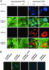
Processing of HBV particles by endosomal lysate enables infection of nonpermissive cells. (A) Confocal microscopy of Huh7 cells infected with unprocessed HBV particles (Left) or with particles incubated with endosomal lysate from HepG2 cells for 30 min before infection (Right). The MGE in both cases was 103. For analysis of the infectivity, HBsAg- and HBcAg-specific antisera were used. Their antigen-specific binding was detected by secondary antibodies visualized by red and blue fluorescence, respectively. Actin filaments were stained by using FITC-conjugated phalloidine. Photographs were taken at ×200 and ×630 magnification. (B) Confocal microscopy of LMH cells infected with unprocessed DHBV particles or with processed WT DHBV, DHBVD1/2, and DHBVD2 particles that had been preincubated with endosomal lysates from LMH cells for 60 min. As a control, processing of WT DHBV was performed in the presence of a protease inhibitor mixture (Roche). The MGE in all cases was 103. For analysis of the infectivity, a surface-specific serum visualized by the blue fluorescence was used. Photographs were taken at ×630 magnification.
Fig. 5.
Fig. 5.
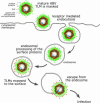
Model of the endosomal processing of hepadnaviral particles. Hepadnaviruses are internalized by receptor-mediated endocytosis. In the endosomal compartment proteolytic cleavage of the surface protein occurs, resulting in a conformational change that exposes the TLMs (shown as red circles) on the surface of the viral particle. The high density of TLMs exposed on the surface of the particle allows endosomal escape into the cytosol to initiate infection.

Similar articles

Cited by

References

    1. Beasley R. P., Hwang L. Y., Lin C. C., Chien C. S. Lancet. 1981;2:1129–1133. - PubMed
    1. Buendia M. A. Semin. Cancer Biol. 2000;10:185–200. - PubMed
    1. Tuttleman J. S., Pugh J. C., Summers J. W. J. Virol. 1986;58:17–25. - PMC - PubMed
    1. Pugh J. C., Summers J. W. Virology. 1989;172:564–572. - PubMed
    1. Glebe D., Aliakbari M., Krass P., Knoop E. V., Valerius K. P., Gerlich W. H. J. Virol. 2003;77:9511–9521. - PMC - PubMed

Publication types

MeSH terms