. 2006 Jun;12(6):1050-60.
doi: 10.1261/rna.3406. Epub 2006 Apr 17.

Interactions of mRNAs and gRNAs involved in trypanosome mitochondrial RNA editing: structure probing of a gRNA bound to its cognate mRNA

Affiliations

Interactions of mRNAs and gRNAs involved in trypanosome mitochondrial RNA editing: structure probing of a gRNA bound to its cognate mRNA

Laura E Yu et al. RNA. 2006 Jun.

Abstract

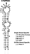
Expression of mitochondrial genes in Trypanosoma brucei requires RNA editing of its mRNA transcripts. During editing, uridylates are precisely inserted and deleted as directed by the gRNA template to create the protein open reading frame. This process involves the bimolecular interaction of the gRNA with its cognate pre-edited mRNA and the assembly of a protein complex with the enzymatic machinery required. While a considerable amount of work has been done identifying the protein components of the editing complex, very little is known about how a functional editosome is assembled. In addition, the importance of RNA structure in establishing a functional editing complex is poorly understood. Work in our lab suggests that different mRNA/gRNA pairs can form similar secondary structures suggesting that a common core architecture may be important for editosome recognition and function. Using solution structure probing, we have investigated the structure of the initiating gRNA, gCYb-558, in the mRNA/gRNA complex with pre-edited apocytochrome b mRNA. Our data indicate that the stem-loop formed by the guiding region of the gRNA alone is maintained in its interaction with the pre-edited message. In addition, our data suggest that a gRNA stem-loop structure is maintained through the first few editing events by the use of alternative base-pairing with the U-tail.


Figures

FIGURE 1.
FIGURE 1.
The predicted models for mRNA/gRNA complex secondary structure have three helices: an mRNA/gRNA anchor helix, a gRNA stem–loop within the guiding region, and a U-tail/mRNA duplex. (A) CYbU-NgCYb-558 pair with the first editing site just 5′ (mRNA) of the anchor indicated by ES1. (B) CYbPES3-NgCYb-558 pair with the fourth editing site just 5′ (mRNA) of the anchor indicated by ES4.
FIGURE 2.
FIGURE 2.
Sequence and alignment of the gRNA/mRNA substrate pairs. (A) The full CYbU (U, unedited) transcript and CYbU aligned to NgCYb-558 is shown. CYbU is composed of 88 nt from the 5′ end of the apocytochrome b mRNA (beginning at +13), including all 13 editing sites as well as a 24-nt vector tag at the 3′ end. (B) The CYbPES3 (PES, partially edited through site 3) transcript and CYbPES3 aligned to NgCYb-558 is shown. The partially edited mRNA, CYbPES3, is identical to CYbU except for the six uridines (lowercase U's) added so that the first three editing sites are fully edited. The complementarity between the gRNA and mRNA in the anchor duplex (bold) is shown with (#) mismatched sequence, (:) GU base pair, (|) Watson–Crick base-pairing. The arrow indicates the position of the introduced cross-link. (C) The NgCYb-558 sequence aligned with wild type initiating CYb gRNAs, gCYb-558, and gCYb[560A] sequences. NgCYb-558 is 59 nt long, including a 15-nt uridylate-tail (U-tail) and has 4 nt differences compared to wild-type gCYb-558. (*) matching sequence, (-) gap. C25, according to NgCYb-558, is bold and underlined in all the gRNAs.
FIGURE 3.
FIGURE 3.
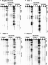
Structure probing of the gRNA, NgCYb-558. Each solution structure probing gel has three sections showing the digestion pattern of NgCYb-558 alone, NgCYb-558 cross-linked to CYbU (+CYbU), and NgCYb-558 cross-linked to CYbPES3 (+CYbPES3). Digestion times in minutes are shown at the top. RNase T1 and U2 ladders with nucleotide numbers are indicated. A line diagram indicating the anchor (bold line), extended anchor (double line), guiding region (thin line), and U-tail of the gRNA is shown next to each digestion pattern. (A) Mung bean nuclease gel, single-strand specific. (B) RNase T2, single-strand specific. (C) RNase T1, single-strand guanosine specific. (D) RNase V1, double-strand specific.
FIGURE 4.
FIGURE 4.
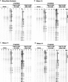
Structure probing of the partially edited CYb mRNA, CYbPES3. Each solution structure probing gel has three sections showing the digestion pattern of CYbPES3 alone, CYbPES3 cross-linked to NgCYb-558sU (no U-tail), and CYbPES3 cross-linked to NgCYb-558. Digestion times in minutes are shown at the top. RNase T1 and U2 ladders with nucleotide numbers are indicated. A diagram of the mRNA is next to each digestion pattern showing the orientation 3′ to 5′ with the extended anchor (bold line). (A) Mung bean nuclease gel, single-strand specific. (B) RNase T2, single-strand specific. (C) RNase T1, single-strand guanosine specific. (D) RNase V1, double-strand specific.
FIGURE 5.
FIGURE 5.
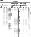
Chemical structure probing of the partially edited CYb mRNA, CYbPES3. The diethyl pyrocarbonate (DEPC) chemical structure probing gel has three sections showing the difference in digestion pattern of CYbPES3 alone, CYbPES3 cross-linked to NgCYb-558sU (no U-tail), and CYbPES3 cross-linked to NgCYb-558. Digestion times in minutes are shown at the top. RNase T1 and U2 ladders with nucleotide numbers are indicated; an alkaline hydrolysis ladder (-OH) was included as well. A diagram of the mRNA is next to each digestion pattern showing the orientation 3′ to 5′ with the extended anchor (bold line).
FIGURE 6.
FIGURE 6.
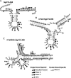
Summary figures from solution structure probing gels that show predicted secondary structures with cleavages from all four nucleases and DEPC chemical. Three sizes of symbols indicate the intensity of cleavage. Circles indicate mung bean nuclease (single-strand specific) cleavages, squares indicate RNase T2 (single-strand specific) cleavages, crosses indicate RNase T1 (single-strand guanosine specific) cleavages, cylinders indicate DEPC (single-strand adenosine specific), and triangles indicate RNase V1 (double-strand specific) cleavages. (A) NgCYb-558 alone. (B) CYbU/NgCYb-558 complex. (C) CYbPES3/NgCYb-558 complex.
FIGURE 7.
FIGURE 7.
Summary figure of CYbPES3 alone from solution structure probing gels that show predicted secondary structure with cleavages from all four nucleases and DEPC using three sizes of symbols to indicate intensity of cleavage. Circles indicate mung bean nuclease (single-strand specific) cleavages, squares indicate RNase T2 (single-strand specific) cleavages, crosses indicate RNase T1 (single-strand guanosine specific) cleavages, cylinders indicate DEPC (single-strand adenosine specific), and triangles indicate RNase V1 (double-strand specific) cleavages.

Similar articles

Cited by

References

    1. Adler B.K., Hajduk S.L. Guide RNA requirement for editing-site-specific endonucleolytic cleavage of preedited mRNA by mitochondrial ribonucleoprotein particles in Trypanosoma brucei . Mol. Cell. Biol. 1997;17:5377–5385. - PMC - PubMed
    1. Aphasizhev R., Simpson L. Isolation and characterization of a U-specific 3′-5′-exonuclease from mitochondria of Leishmania tarentolae . J. Biol. Chem. 2001;276:21280–21284. - PubMed
    1. Aphasizhev R., Sbicego S., Peris M., Jang S.H., Aphasizheva I., Simpson A.M., Rivlin A., Simpson L. Trypanosome mitochondrial 3′ terminal uridylyl transferase (TUTase): The key enzyme in U-insertion/deletion RNA editing. Cell. 2002;108:637–648. - PubMed
    1. Aphasizhev R., Aphasizheva I., Nelson R.E., Gao G., Simpson A.M., Kang X., Falick A.M., Sbicego S., Simpson L. Isolation of a U-insertion/deletion editing complex from Leishmania tarentolae mitochondria. EMBO J. 2003a;22:913–924. - PMC - PubMed
    1. Aphasizhev R., Aphasizheva I., Nelson R.E., Simpson L. A 100-kD complex of two RNA-binding proteins from mitochondria of Leishmania tarentolae catalyzes RNA annealing and interacts with several RNA editing components. RNA. 2003b;9:62–76. - PMC - PubMed

Publication types

MeSH terms

LinkOut - more resources