. 2006 Jun;12(6):968-79.
doi: 10.1261/rna.50506. Epub 2006 Apr 17.

Arrested yeast splicing complexes indicate stepwise snRNP recruitment during in vivo spliceosome assembly

Affiliations

Arrested yeast splicing complexes indicate stepwise snRNP recruitment during in vivo spliceosome assembly

Daniel F Tardiff et al. RNA. 2006 Jun.

Abstract

Pre-mRNA splicing is catalyzed by the spliceosome, a macromolecular machine dedicated to intron removal and exon ligation. Despite an abundance of in vitro information and a small number of in vivo studies, the pathway of yeast (Saccharomyces cerevisiae) in vivo spliceosome assembly remains uncertain. To address this situation, we combined in vivo depletions of U1, U2, or U5 snRNAs with chromatin immunoprecipitation (ChIP) analysis of other splicing snRNPs along an intron-containing gene. The data indicate that snRNP recruitment to nascent pre-mRNA predominantly proceeds via the canonical three-step assembly pathway: first U1, then U2, and finally the U4/U6*U5 tri-snRNP. Tandem affinity purification (TAP) using a U2 snRNP-tagged protein allowed the characterization of in vivo assembled higher-order splicing complexes. Consistent with an independent snRNP assembly pathway, we observed high levels of U1-U2 prespliceosomes under U5-depletion conditions, and we observed significant levels of a U2/U5/U6/Prp19-complex mature splicing complex under wild-type conditions. These complexes have implications for the steady-state distribution of snRNPs within nuclei and also reinforce the stepwise recruitment of U1, U2, and the tri-snRNP during in vivo spliceosome assembly.


Figures
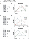
FIGURE 1.
FIGURE 1.
U snRNA depletions affect cotranscriptional recruitment of U1, U2, and U5 snRNPs. TAP-tagged U snRNA depletion strains were grown in galactose or glucose prior to chromatin immunoprecipitations (ChIPs). Primer extensions confirming depletion of snRNAs and inhibition of splicing (as evident by accumulation of pre-U3a/b snRNA) are shown in the left panels (B,D,F). For ChIP (C,E,G), primer sets spanning the ACT1 gene (A) were used to monitor U snRNP association. The Y-axis reflects enrichment of U snRNPs to ACT1 relative to the intronless PMA1 gene to control for background binding. The X-axis represents distance from the ACT1 start codon. The black bar demarcates the location of the intron. The snRNP factors immunoprecipitated and found in graph legends are the following: U1 (U1C-TAP), U2 (Lea1p-TAP), and U5 (Prp8p-TAP). (B,C) U1 snRNA depletion, (D,E) U2 snRNA depletion, and (F,G) U5 snRNA depletion.
FIGURE 2.
FIGURE 2.
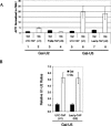
Post-transcriptional splicing complexes accumulate in snRNA depletions. (A) The Gal-U2 and Gal-U5 TAP tagged strains described in Figure 1 were used in a U snRNP immunoprecipitation experiment (see Materials and Methods). Values represent ACT1 levels normalized to intronless PMA1 to show enrichment over background. Black columns indicate strains grown in galactose (nondepletion), and white columns indicate those grown in glucose (depletion). ACT1 RNA is enriched on U1 snRNP (U1C-TAP) in the absence of U2 snRNP while U5 snRNP (Prp8p-TAP) levels decrease. Increased association of ACT1 with U1 indicates accumulation of commitment complexes. In U5-depletion strains, ACT1 is highly enriched on both U1 (U1C-TAP) and U2 snRNPs (Lea1p-TAP), indicative of prespliceosome formation. (B) U1 and U2 snRNAs from the same U snRNP IP experiments were analyzed by quantitative RT-PCR in Gal-U5 U1C-TAP and LEA1-TAP. Black columns represent the U1–U2 ratios when grown in galactose; white columns indicate the U1–U2 ratios in U5 depletion. In U1C-TAP, the ratio is U2:U1 and for LEA1-TAP, the ratio is U1:U2. U2 is more highly associated with U1 in Gal–U5 U1C-TAP and U1 more highly associated with U2 in Gal–U5 LEA1-TAP upon depletion. The reciprocal increased association between U1 and U2 suggests prespliceosomes form in the absence of U5.
FIGURE 3.
FIGURE 3.
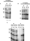
Characterization of U2 snRNP-associated U snRNAs (A). U snRNAs from Gal–U5 LEA1-TAP were analyzed by primer extension following TAP purification from cells grown in galactose and glucose. In galactose, U2 is most highly associated with U5 and U6 snRNAs (low levels of U1 and U4 snRNA). In U5 depletion, U1, U5, and U6 snRNAs associate with U2 snRNP. (B) Lea1p-TAP immunoprecipitates predominantly U2 snRNA upon U1 depletion, indicating that U5 and U6 snRNP association is splicing dependent. (C) Extracts generated via traditional French press methods or our modified protocol were compared for association of snRNAs with Lea1p-TAP. FP, French press; BB, bead beating. LEA1-TAP or an untagged wild-type strain were used for immunoprecipitations. Lea1p-TAP specifically immunoprecipitates U2 snRNA when using traditional methods. However, with modified conditions, Lea1p-TAP interacts most highly with U2, U5, and U6 snRNAs with lower levels of U1 and U4.
FIGURE 4.
FIGURE 4.
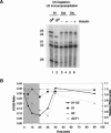
Dynamics of prespliceosomes and U2/U5/U6 complexes during thiolutin treatment. (A) U snRNAs from Gal-U5 LEA1-TAP were analyzed by primer extension following TAP purification from cells grown in galactose and glucose with or without thiolutin (see Materials and Methods). Only U1–U2 prespliceosomes in U5 depletion turn over with thiolutin treatment, indicating that there are two separate complexes associated with U2 snRNP. (B) Prespliceosomes were monitored during thiolutin treatment and return to growth in Gal–U5 LEA1-TAP (see Materials and Methods). The gray section indicates thiolutin treatment; the white indicates time after thiolutin removal. The primary Y-axis represents U1–U2 ratios (prespliceosomes), and the secondary Y-axis represents U1, U2, and ACT1 mRNA all normalized to U2 snRNA levels and then to the first time point. Prespliceosomes decay with a half-life of ∼5 min and form with similar kinetics upon thiolutin removal and initiation of transcription. U1, U2, and ACT1 RNAs were analyzed from the same experiment to confirm effectiveness of thiolutin treatment. U1 and U2 snRNAs are stable during the thiolutin treatment, while ACT1 is labile and recovers with removal of thiolutin.
FIGURE 5.
FIGURE 5.
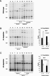
High levels of U2 snRNP are in higher-order splicing complexes in vivo. Sucrose gradients of wild-type (A), U1-depleted (B), and U5-depleted (C) cells are shown. Extracts were layered onto 10%–30% sucrose gradients and fractions analyzed by primer extension. U snRNA distribution was quantitated from at least two independent sucrose gradients and is shown to the right of the corresponding gradient (see Materials and Methods). After U1 depletion (cf. B and A), a considerable fraction of U2 snRNP migrates as a mono-snRNP, indicating that under normal physiological conditions, a major fraction (∼50%) of U2 snRNP is in higher-order complexes. After U5 depletion (cf. C and A), U1 snRNA redistributes across the gradient toward larger complexes, consistent with accumulation of U1–U2 prespliceosomes.
FIGURE 6.
FIGURE 6.
Spliceosome assembly in U snRNA depletion strains. Spliceosome assembly pathways are shown in Gal–U1, Gal–U2, Gal–U5, and wild-type (WT) strains. Cotranscriptional and post-transcriptional complexes (circled) are depicted. No spliceosome assembly occurs in the absence of U1 snRNP (Gal–U1). However, commitment complex forms cotranscriptionally upon U2 snRNP depletion, while tri-snRNP addition is inhibited (Gal–U2). In the absence of U5 snRNP (tri-snRNP), prespliceosomes are only observed post-transcriptionally because free U1 and U2 are titrated away from the site of transcription (Gal–U5). Our data suggest that spliceosome assembly proceeds in a stepwise fashion in vivo and does not require higher-order snRNP complexes (tetra-snRNP, penta-snRNP, etc.).

Similar articles

Cited by

References

    1. Abruzzi K.C., Lacadie S., Rosbash M. Biochemical analysis of TREX complex recruitment to intronless and intron-containing yeast genes. EMBO J. 2004;23:2620–2631. - PMC - PubMed
    1. Bouveret E., Rigaut G., Shevchenko A., Wilm M., Seraphin B. A Sm-like protein complex that participates in mRNA degradation. EMBO J. 2000;19:1661–1671. - PMC - PubMed
    1. Burge C.B., Tuschl T., Sharp P.A. Splicing of precursors to mRNAs by the spliceosome. In: Gesteland R.R., et al., editors. The RNA world. 2d ed. Cold Spring Harbor Laboratory Press; Cold Spring Harbor, NY: 1999. pp. 525–560.
    1. Caspary F., Shevchenko A., Wilm M., Seraphin B. Partial purification of the yeast U2 snRNP reveals a novel yeast pre- mRNA splicing factor required for pre-spliceosome assembly. EMBO J. 1999;18:3463–3474. - PMC - PubMed
    1. Das R., Zhou Z., Reed R. Functional association of U2 snRNP with the ATP-independent spliceosomal complex E. Mol. Cell. 2000;5:779–787. - PubMed

Publication types

MeSH terms

Substances