. 2006 May 3;25(9):1827-35.
doi: 10.1038/sj.emboj.7601088. Epub 2006 Apr 13.

The Hrd1p ligase complex forms a linchpin between ER-lumenal substrate selection and Cdc48p recruitment

Affiliations

The Hrd1p ligase complex forms a linchpin between ER-lumenal substrate selection and Cdc48p recruitment

Robert Gauss et al. EMBO J. .

Abstract

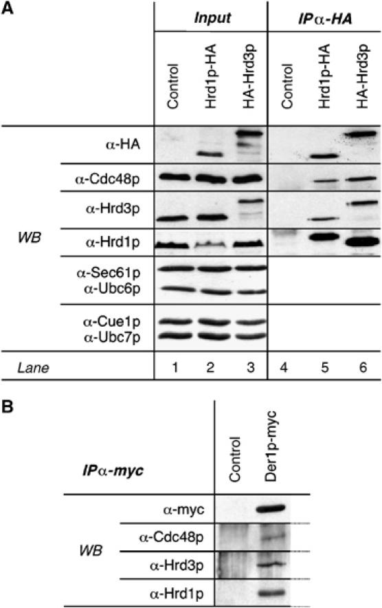
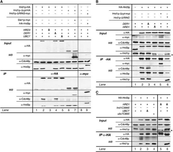
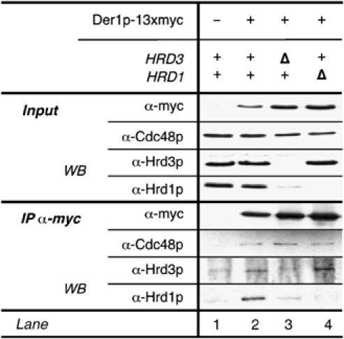
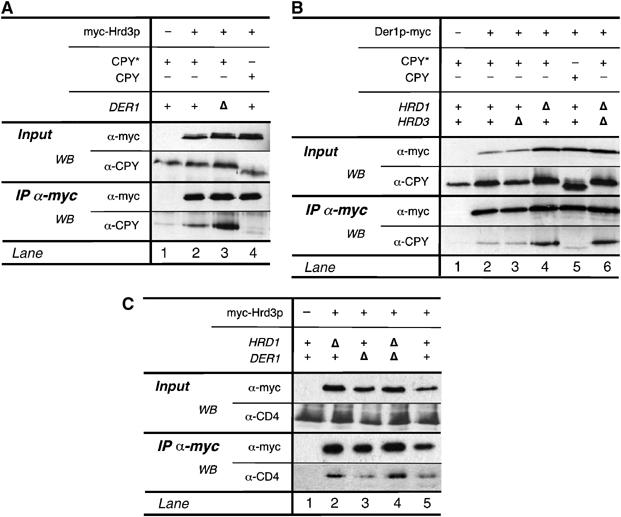
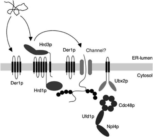
Misfolded proteins of the endoplasmic reticulum (ER) are targeted to the cytoplasm for proteasomal degradation. Key components of this process are ER membrane-bound ubiquitin ligases. These ligases associate with the cytoplasmic AAA-ATPase Cdc48p/p97, which is thought to support the release of malfolded proteins from the ER. Here, we characterize a yeast protein complex containing the ubiquitin ligase Hrd1p and the ER membrane proteins Hrd3p and Der1p. Hrd3p binds malfolded proteins in the ER lumen enabling their delivery to downstream components. Therefore, we propose that Hrd3p acts as a substrate recruitment factor for the Hrd1p ligase complex. Hrd3p function is also required for the association of Cdc48p with Hrd1p. Moreover, our data demonstrate that recruitment of Cdc48p depends on substrate processing by the Hrd1p ligase complex. Thus, the Hrd1p ligase complex unites substrate selection in the ER lumen and polyubiquitination in the cytoplasm and links these processes to the release of ER proteins via the Cdc48p complex.


Figures

Figure 1
Figure 1
Hrd1p, Hrd3p, and Der1p form a heterogenic complex and associate with cytosolic Cdc48p. (A) Microsomes were isolated from logarithmically growing cells expressing Hrd1p-HA or HA-Hrd3p (see Materials and methods). Subsequently, membranes were solubilized with NP40 using nondenaturing conditions and HA-tagged proteins were immunoprecipitated. Precipitates were analyzed by SDS–PAGE and immunoblotting using specific antibodies. The Hrd1p antiserum reacts only poorly with the C-terminally HA-tagged Hrd1p protein (see also Supplementary Figure 1). The signal in lane 2 is an unspecific crossreaction of the Hrd1p antiserum. (B) Der1p-myc was immunoprecipitated from isolated microsomes as in (A) and the precipitate analyzed via immunoblotting.
Figure 2
Figure 2
Hrd3p and the catalytic activity of Hrd1p are essential for the recruitment of Cdc48p. (A) Hrd1p-HA and epitope-tagged mutant forms of Hrd1p lacking either the cytoplasmic C-terminus (Hrd1p-Δcyt) or the RING-domain (Hrd1p-ΔRING; for description of the mutants, see Materials and methods section) were immunoprecipitated from solubilized membrane preparations of the indicated yeast strains as described in Figure 1. Precipitated material was subsequently analyzed by immunoblotting. (B, C) HA-Hrd3p was immunoprecipitated from the indicated yeast strains. Note that the Hrd1p polyclonal antibody was raised against a region not present in Hrd1p-Δcyt and hence this serum does not react with Hrd1p-Δcyt. (*) Refers to unspecific cross-reacting material. (▵) Indicates deletion of the respective gene.
Figure 3
Figure 3
Der1p binds to the Hrd1p complex and interacts with Cdc48p. An myc-tagged version of Der1p as immunoprecipitated from isolated membrane preparations of the indicated yeast strains under nondenaturing conditions and co-precipitating proteins were identified via immunoblotting.
Figure 4
Figure 4
Hrd3p and Der1p autonomously bind the misfolded protein CPY*. myc-Hrd3p (A) and Der1p-myc (B) were precipitated from the indicated yeast strains expressing CPY* or wild-type CPY. Co-precipitated material was analyzed by Western blotting. (C) CD4 interacts with Hrd3p. myc-Hrd3p was immunopurified from yeast cells heterologously expressing CD4. The relevant genotype of the yeast strains used is given.
Figure 5
Figure 5
Hrd3p and Der1p are capable of recruiting CPY* to the Hrd1p ligase. Hrd1p-HA and Hrd1p-Δcyt-HA were pulled down from solubilized membrane fractions isolated from the indicated yeast strains. Bound CPY* or CPY was detected by immunoblotting using a specific antibody.
Figure 6
Figure 6
The C-terminal part of the ER-lumenal domain of Hrd3p is essential to trigger efficient degradation of CPY*. (A) Schematic domain structure of Hrd3p with representation of C-terminal truncation variants and a summary of the results. (B) A C-terminal region within the ER-lumenal domain of Hrd3p is essential for efficient CPY* degradation. Strains expressing wild-type Hrd3p and the various truncation mutants were essayed for CPY* degradation by pulse-chase analysis. Displayed is a representative experiment. (C) Quantitative evaluation of three pulse-chase experiments with error bars indicating the standard deviation of mean values.
Figure 7
Figure 7
Hrd3p connects ER-lumenal substrate recognition and cytosolic Cdc48p binding. HA-Hrd3p and HA-tagged truncations mutants (Hrd3p-Δ801, -Δ769, and -Δ664) were pull-down from solubilized microsomal preparations. Precipitates were analyzed for Hrd3p-bound Cdc48, Hrd1p, and CPY* by immunoblot using specific antibodies.
Figure 8
Figure 8
The Hrd1p ligase complex provides a linchpin between substrate selection in the ER and the binding of Cdc48p in the cytoplasm. Misfolded ER proteins bind to the ER-luminal domain of Hrd3p and possibly to Der1p. Subsequently, these substrates are dislocated into the cytoplasm. It is still a matter of debate how these proteins pass the ER membrane. Der1p as well as membrane-bound ubiquitin ligases such as Hrd1p or Doa10p may participate in this process. During or after dislocation, the substrates are ubiquitinated by Hrd1p. In the cytoplasm, binding of Cdc48p–Npl4p–Ufd1p supports the dislocation of ER proteins and may also target them to the proteasome. Recruitment of the Cdc48p complex to Hrd1p depends on the membrane protein Ubx2p/Sel1p as well as the catalytic activity of the ubiquitin ligases.

References

    1. Ausubel FM (eds) (1993–2004) Current Protocols in Molecular Biology. New York: John Wiley & Sons, Inc.
    1. Bays NW, Gardner RG, Seelig LP, Joazeiro CA, Hampton RY (2001a) Hrd1p/Der3p is a membrane-anchored ubiquitin ligase required for ER-associated degradation. Nat Cell Biol 3: 24–29 - PubMed
    1. Bays NW, Wilhovsky SK, Goradia A, Hodgkiss-Harlow K, Hampton RY (2001b) HRD4/NPL4 is required for the proteasomal processing of ubiquitinated ER proteins. Mol Biol Cell 12: 4114–4128 - PMC - PubMed
    1. Biederer T, Volkwein C, Sommer T (1996) Degradation of subunits of the Sec61p complex, an integral component of the ER membrane, by the ubiquitin–proteasome pathway. EMBO J 15: 2069–2076 - PMC - PubMed
    1. Biederer T, Volkwein C, Sommer T (1997) Role of Cue1p in ubiquitination and degradation at the ER surface. Science 278: 1806–1809 - PubMed

Publication types

MeSH terms