. 2006 May 3;25(9):1957-66.
doi: 10.1038/sj.emboj.7601087. Epub 2006 Apr 13.

Definition of the bacterial N-glycosylation site consensus sequence

Affiliations

Definition of the bacterial N-glycosylation site consensus sequence

Michael Kowarik et al. EMBO J. .

Abstract

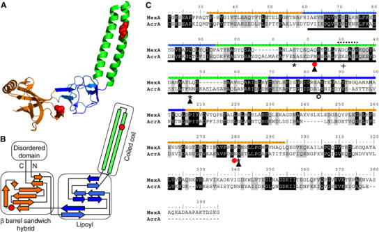
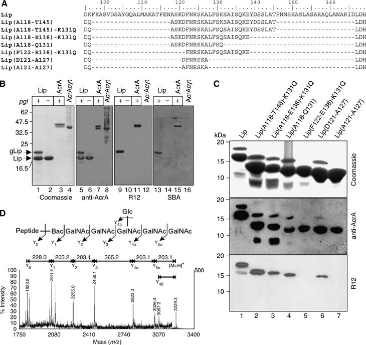
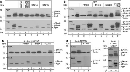
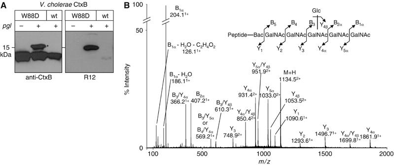
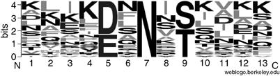
The Campylobacter jejuni pgl locus encodes an N-linked protein glycosylation machinery that can be functionally transferred into Escherichia coli. In this system, we analyzed the elements in the C. jejuni N-glycoprotein AcrA required for accepting an N-glycan. We found that the eukaryotic primary consensus sequence for N-glycosylation is N terminally extended to D/E-Y-N-X-S/T (Y, X not equalP) for recognition by the bacterial oligosaccharyltransferase (OST) PglB. However, not all consensus sequences were N-glycosylated when they were either artificially introduced or when they were present in non-C. jejuni proteins. We were able to produce recombinant glycoproteins with engineered N-glycosylation sites and confirmed the requirement for a negatively charged side chain at position -2 in C. jejuni N-glycoproteins. N-glycosylation of AcrA by the eukaryotic OST in Saccharomyces cerevisiae occurred independent of the acidic residue at the -2 position. Thus, bacterial N-glycosylation site selection is more specific than the eukaryotic equivalent with respect to the polypeptide acceptor sequence.


Figures

Figure 1
Figure 1
The structural homology of MexA and AcrA. (A) Crystal structure of the MexA protein (adapted from Higgins et al, 2004). Colors indicate the different domains. Red spheres show a space fill representation of the residues in MexA corresponding to the glycosylated N123 and N273 of AcrA based on the homology alignment (C). (B) Scheme of the secondary structure elements in MexA. Orange is the β barrel sandwich hybrid domain. It is connected to an unordered part of the protein containing the N and C termini. Blue: lipoyl domain. Green: coiled coil. Note the overall antiparallel organization of the polypeptide. Red circles indicate the residues in MexA, which align to N123 and N273 (C). (C) Polypeptide alignment of signal peptide processed MexA and AcrA amino acid sequences. The ruler is adjusted to the AcrA amino acid numbering. Colored lines indicate the sequences constituting the domains in MexA (see A, B). The dashed line designates the loop connecting the ascending and descending strands of the coiled coil in MexA. The black line below indicates the part of AcrA constituting the Lip sequence (see Figure 2); the red circles show the naturally glycosylated N123 and N273 of AcrA, the asterisk and the empty circle discriminate Asn residues in additionally introduced glycosylation sites that were active (N117, N145, N274) or inactive (N184, see Figure 3E). Filled triangles indicate glycosylation sites, which were glycosylated in the yeast expression system (see Figure 6). + shows the residue in the protease sensitive loop that was replaced by Gln (K131Q, see Figure 3). Black and gray shading shows identical and similar amino acid residues according to the BLOSUM62 substitution matrix. Note that the AcrA sequence corresponding to the coiled coil is longer than the homologous stretch of MexA.
Figure 2
Figure 2
Glycosylation analysis of truncated forms of AcrA. (A) Sequence details of the hypothetical coiled coil domain (see Figure 1, the green domain) present in Lip and truncated forms thereof, which were tested as N-glycan acceptors in E. coli. The ruler indicates the amino acid numbering of the corresponding residues as in the AcrA sequence. Residues D95 and L167 belong to the predicted lipoyl domain and flank the sequence of the coiled coil that include the glycosylation at N123. The peptide stretches of the different Lip truncation variants containing the glycosylation site at N123 are indicated in brackets, according to the numbering in the AcrA sequence. According to this nomenclature, the full length, nontruncated Lip protein is Lip(K96-D166). Note that some residues were mutated due to cloning reasons or to render the protein more resistant to proteolytic cleavage (K131Q). (B) Characterization of the glycosylated lipoyl coiled coil domain of AcrA (Lip). SDS–PAGE analysis of four different Ni2+ affinity purified protein fractions by Coomassie Brilliant Blue staining (left panel), immunoblotting with anti-AcrA antiserum (middle left), R12 antiserum (middle right), or HRP-coupled SBA (right panel). Lip was expressed in presence (+, lanes 1, 5, 9, and 13) and absence (−, lanes 2, 6, 10, and 14) of a functional pgl locus, soluble AcrA only in its presence (+, lanes 3, 7, 11, and 15). Unglycosylated AcrA was purified from the cytoplasm (lanes 4, 8, 12, and 16; Nita-Lazar et al, 2005). Note that the glycosylated proteins are detected with the R12 antiserum and SBA. (C) Glycosylation analysis of truncated forms of Lip. Proteins were Ni2+ affinity purified from periplasmic extracts of Top10 cells expressing the corresponding proteins as indicated above the gel frame and analyzed by SDS–PAGE and immunoblotting. Top gel: Coomassie stained; middle gel: anti-AcrA; bottom: R12. (D) MALDI-MS/MS of m/z=3229.3 from the in-gel trypsinized protein band labeled with a star in panel C. The mass corresponds to the expected glycopeptide derived from the Lip(D121-A127) protein (GQTLFIIEQDQDFN123R). The inset shows the C. jejuni N-glycan attached to the peptide and the corresponding fragmentation pattern.
Figure 3
Figure 3
SDS–PAGE analysis of N-glycosylation site mutants of AcrA. All top panels are immunoblots probed with anti-AcrA antiserum, bottom panels represent identical samples detected with the C. jejuni N-glycan specific R12 antiserum. + and − indicate the presence of pACYCpgl or pACYCpglmut in the cells. All samples contained a plasmid expressing soluble AcrA with an N-terminal signal peptide. The different point mutations in the AcrA protein are indicated. Numbers on the left of the gel frame show the electrophoretic mobility of the molecular weight marker. The numbers of N-glycans in the different glycoforms of AcrA are indicated at the right of the gel frames. (A) Analysis of the naturally used glycosylation sites N123 and N273. (B) Analysis of artificially introduced glycosylation sites at positions N117, N147, and N274, in the presence of active natural glycosylation sites. (C) Analysis of artificially introduced glycosylation sites at positions N117, N147, and N274, in the absence of other glycosylation sites. (D) Pro in position −1 of the natural glycosylated N123 abolishes glycosylation. (E) A sequon that was mutated into the putative lipoyl domain of AcrA was not active.
Figure 4
Figure 4
Bacterial N-Glycosylation of a non-C. jejuni protein. (A) Periplasmic extracts derived from cells expressing wild-type CtxB and CtxB-W88D were analyzed by immunoblotting with anti-CtxB antiserum (left panel) and R12 (right panel). Expression was performed in SCM7 E. coli cells containing either the glycosylation competent (+) or incompetent (−) pgl machinery on a plasmid. (B) Ni2+ purified, in-gel trypsinized protein corresponding to the glycosylated CtxB-W88D (marked with an asterisk in (A)) was analyzed by NanoESI-MS/MS. The fragmentation spectrum of the doubly charged pseudomolecular ion at m/z=1134.5, corresponding to the tryptic CtxB glycopeptide bearing the C. jejuni N-glycan is shown. The inset illustrates the C. jejuni N-glycan attached to the expected peptide, with sequential losses of HexNAc (203 Da) and Hex (162 Da) residues confirming the expected structure.
Figure 5
Figure 5
Statistical analysis of active N-glycosylation sites found in native C.jejuni glycoproteins. Fractional occurrence of amino acids in the region from −6 to +6 of the glycosylated Asn residue (32 sequons). The figure was prepared from the data of Table I, by means of WebLogo (Crooks et al, 2004). A full color version of this figure is provided in Supplementary Figure S4.
Figure 6
Figure 6
AcrA N-glycosylation site usage in yeast. AcrA was expressed in yeast cells with an N-terminal signal sequence for secretion. Shown are ConA lectin-purified fractions from cells expressing either wild-type AcrA or glycosylation site point mutants as indicated above the gel frame to analyze the site usage. EndoH treatment (+) was performed to show that all three bands obtained with wild-type AcrA contained one or more high-mannose N-glycans.

References

    1. Akama H, Matsuura T, Kashiwagi S, Yoneyama H, Narita S, Tsukihara T, Nakagawa A, Nakae T (2004) Crystal structure of the membrane fusion protein, MexA, of the multidrug transporter in Pseudomonas aeruginosa. J Biol Chem 279: 25939–25942 - PubMed
    1. Alaimo C, Catrein I, Morf L, Marolda CL, Callewaert N, Valvano MA, Feldman MF, Aebi M (2006) Two distinct but interchangeable mechanisms for flipping of lipid-linked oligosaccharides. EMBO J 25: 967–976 - PMC - PubMed
    1. Bause E, Breuer W, Peters S (1995) Investigation of the active site of oligosaccharyltransferase from pig liver using synthetic tripeptides as tools. Biochem J 312 (Part 3): 979–985 - PMC - PubMed
    1. Ben-Dor S, Esterman N, Rubin E, Sharon N (2004) Biases and complex patterns in the residues flanking protein N-glycosylation sites. Glycobiology 14: 95–101 - PubMed
    1. Bulleid NJ, Bassel-Duby RS, Freedman RB, Sambrook JF, Gething MJ (1992) Cell-free synthesis of enzymically active tissue-type plasminogen activator. Protein folding determines the extent of N-linked glycosylation. Biochem J 286 (Part 1): 275–280 - PMC - PubMed

Publication types

MeSH terms

LinkOut - more resources