. 2006 May 3;25(9):2007-16.
doi: 10.1038/sj.emboj.7601084. Epub 2006 Apr 13.

Role of SGT1 in resistance protein accumulation in plant immunity

Affiliations

Role of SGT1 in resistance protein accumulation in plant immunity

Cristina Azevedo et al. EMBO J. .

Abstract

A highly conserved eukaryotic protein SGT1 binds specifically to the molecular chaperone, HSP90. In plants, SGT1 positively regulates disease resistance conferred by many Resistance (R) proteins and developmental responses to the phytohormone, auxin. We show that silencing of SGT1 in Nicotiana benthamiana causes a reduction in steady-state levels of the R protein, Rx. These data support a role of SGT1 in R protein accumulation, possibly at the level of complex assembly. In Arabidopsis, two SGT1 proteins, AtSGT1a and AtSGT1b, are functionally redundant early in development. AtSGT1a and AtSGT1b are induced in leaves upon infection and either protein can function in resistance once a certain level is attained, depending on the R protein tested. In unchallenged tissues, steady-state AtSGT1b levels are at least four times greater than AtSGT1a. While the respective tetratricopeptide repeat (TPR) domains of SGT1a and SGT1b control protein accumulation, they are dispensable for intrinsic functions of SGT1 in resistance and auxin responses.


Figures

Figure 1
Figure 1
NbSGT1 positively controls steady-state levels of Rx. Western blot analysis of Rx, NbSGT1 and HSP90 levels using α-HA, α-SGSa and α-HSP90 antibodies, respectively. The Rx-4HA transgenic N. benthamiana plants were inoculated with TRV:00 (control), TRV:NbSGT1 or TRV:NbEDS1. Plants were sampled at 13–16 days postinoculation. Protein extracts were prepared as previously described (Bieri et al, 2004). Equal loading of total protein was checked by Ponceau S staining of Rubisco. For each treatment results, representative of three independent experiments are shown.
Figure 2
Figure 2
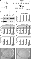
Analysis of of Arabidopsis sgt1a-1 and rar1-1 mutants. (A) Relative position of T-DNA insertions within the AtSGT1a and AtRAR1 genes. Exons are indicated by the black boxes. (B) Protein expression of AtSGT1a, AtSGT1b and AtRAR1. Western blot analysis of total protein extracts from mutant and wild-type plants probed with α-SGSa or α-RAR1 antibody, as indicated. (C) Bacterial growth analysis of Pst DC3000 (vector) after infiltration of leaves of 6–7 week-old plants with bacterial suspensions (1 × 10⁵ c.f.u./ml). Leaves were harvested at 0 (white column) and 3 days (grey column) after inoculation. (D) Same analysis for Pst DC3000 (avrRpm1), (E) Pst DC3000 (avrRpt2), (F) Pst DC3000 (avrRphB), (G) Pst DC3000 (avrRps4). Asterisks indicate that bacterial growth is significantly different (P<0.05) from the wild-type control. Experiments were repeated at least three times with similar results. (H) DIC image of cleared seed from wild-type plant. (I) DIC image of cleared seed from a self-pollinated mutant plant homozygous for sgt1a-1 mutation and heterozygous for sgt1b-1.
Figure 3
Figure 3
AtSGT1a and AtSGT1b expression is induced by pathogen infection. Induction of β-glucuronidase (GUS) activity in Ler transgenics expressing pAtSGT1a∷GUS (A, C, E) or pAtSGT1b∷GUS (B, D, F) was examined 3 and 7 days after inoculation (dpi) with avirulent (Noco2) or virulent (Cala2) H. parasitica, as indicated. GUS-stained leaves were viewed under a light microscope. Pictures are representative of three independent experiments using three independent transgenic lines for each construct. HR, hypersensitive response at pathogen infection foci; M, pathogen mycelium; O; pathogen oospores; V, vasculature. Bars represent 200 μm.
Figure 4
Figure 4
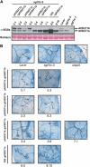
Expression of AtSGT1a or AtSGT1b in transgenic Arabidopsis complements sgt1b-3 in RPP5 resistance. (A) Western blot analysis of total leaf extracts from different sgt1b-3 plants transformed with AtSGT1a or AtSGT1b under different promoters. SGT1 proteins were visualized by the α-SGSa antibody. (B) Lactophenol trypan blue staining of Noco-2 infected leaves 7 days after inoculation reveals necrotic plant cells and pathogen structures. HR, hypersensitive response; M, mycelium; TN, trailing plant cell necrosis; S, sporangiophore. Bars represent 500 μm.
Figure 5
Figure 5
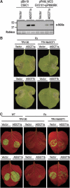
AtSGT1a and AtSGT1b are functional in Rx resistance. (A) Western blot analysis of differential AtSGT1a protein expression levels by Agrobacterium strains C58C1 with a pBIN61-based vector and GV3101 with a pPAM-MCS vector. Protein extracts from N. benthamiana leaves infiltrated with indicated strains were separated by SDS–PAGE and blotted on a membrane. AtSGT1a was visualized by α-SGSa antibody. (B) Appearance of HR elicited by co-infiltration of Agrobacterium expressing the PVX-Tk (PVX coat protein) and the test constructs. One week-old wild type and transgenic N. benthamiana plants (Rx, N) were inoculated with TRV:NbSGT1 or TRV:00 by Agrobacterium infiltration. Approximately 3 weeks later PVX elicitor was co-expressed with the test constructs AtSGT1a, AtSGT1b or empty vector by Agrobacterium infiltration. Plants were photographed 5 days postinoculation under white light. (C) Rx resistance against PVX. Co-infiltration of Agrobacterium carrying PVX:GFP (OD600=0.001) and the test construct (OD600=0.25) 3 weeks after inoculation of TRV:00 or TRV:NbSGT1. Accumulation of PVX:GFP was monitored by GFP fluorescence under UV illumination 5 days post inoculation. Similar results were obtained in three independent experiments.
Figure 6
Figure 6
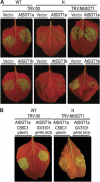
Dose-dependent recruitment of AtSGT1a in N resistance. (A) AtSGT1b but not AtSGT1a is functional in N resistance against TMV when expressed from the Agrobacterium strain GV3101 (pMP90RK) in a pPAM-MCS-based vector. Wild type and transgenic N. benthamiana plants were treated as described in Figure 5. Co-infiltration of Agrobacterium carrying TMV:GFP (OD600=0.025) and the test construct (OD600=0.25) as indicated. Accumulation of TMV:GFP was monitored by GFP fluorescence under UV illumination 6-7 days post inoculation. Similar results were obtained for three independent experiments. (B) Comparison of two Agrobacterium strains, C58C1 with a pBIN61-based vector and GV3101 with a pPAM-MCS vector for AtSGT1a functionality in N resistance.
Figure 7
Figure 7
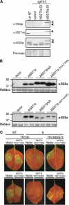
The TPR domain controls steady-state levels of Arabidopsis SGT1 proteins. (A) Western blot analysis of AtSGT1a-SII (indicated by black arrow) and AtSGT1b-SII (white arrow). (B) Relative abundance of different AtSGT1a and AtSGT1b constructs transiently expressed in TRV:NbSGT1 plants determined by Western blot analysis with the α-SGSa antibody. A chimeric construct AtSGT1a/b/b containing the TPR domain (M1-V163) from AtSGT1a and the remaining protein from AtSGT1b (V171-Y357) while the reciprocal chimera AtSGT1b/a/a contains M1-A170 from AtSGT1b fused to V164-I350 of AtSGT1a. (C) AtSGT1a(T91A+T100A) and AtSGT1b(A91T+A100T) contructs were transiently expressed in TRV:00 or TRV:NbSGT1 plants as indicated and analysis of N resistance performed as in Figure 6A. Accumulation of TMV:GFP was monitored by GFP fluorescence under UV illumination 6–7 days post inoculation.
Figure 8
Figure 8
The TPR domain is dispensable for Rx resistance in N. benthamiana. ΔTPRa and ΔTPRb were expressed using the Agrobacterium strain GV3101 (pMP90RK) with a pPAM_MCS-based vector. Experiments were performed as described in Figure 5.
Figure 9
Figure 9
Model for SGT1 function in assembly of plant NB-LRR protein complexes. An NB-LRR type R protein is depicted as part of a ‘preactivated' multiprotein complex with other plant protein(s) (shown as x), whose assembly requires the cooperative activities of the HSP90 chaperone and RAR1. Our data argue for a role of SGT1 in assembly (solid straight black arrow) of pre-existing NB-LRR proteins. In Arabidopsis, two active SGT1 isoforms, AtSGT1a and AtSGT1b, are expressed. The predominant SGT1 activity in leaves is exerted by AtSGT1b due to its higher accumulation (indicated by dark grey shading) than AtSGT1a (light grey shading) before pathogen challenge, and possibly also preferential HSP90 binding. In the absence of AtSGT1b and RAR1, AtSGT1a activity is sufficient for accumulation of some NB-LRR proteins, such as RPS5, but not others, such as RPP5 or RPM1. This function is supported by an intrinsic capability of AtSGT1a to complement the sgt1b mutant when expressed at a certain level and by differences in the amounts of SGT1 needed for resistance conferred by various R proteins tested. It is likely that a fine balance between NB-LRR protein assembly and degradation is mediated by chaperone and co-chaperone associations. While the model depicts a principle activity of SGT1 in R protein assembly, it does not preclude an SGT1 contribution to degradation of R proteins (broken straight black arrow) or downstream signalling components.

References

    1. Austin MJ, Muskett P, Kahn K, Feys BJ, Jones JD, Parker JE (2002) Regulatory role of SGT1 in early R gene-mediated plant defenses. Science 295: 2077–2080 - PubMed
    1. Azevedo C, Sadanandom A, Kitagawa K, Freialdenhoven A, Shirasu K, Schulze-Lefert P (2002) The RAR1 interactor SGT1, an essential component of R gene-triggered disease resistance. Science 295: 2073–2076 - PubMed
    1. Bansal PK, Abdulle R, Kitagawa K (2004) Sgt1 associates with Hsp90: an initial step of assembly of the core kinetochore complex. Mol Cell Biol 24: 8069–8079 - PMC - PubMed
    1. Belkhadir Y, Subramaniam R, Dangl JL (2004) Plant disease resistance protein signaling: NBS-LRR proteins and their partners. Curr Opin Plant Biol 7: 391–399 - PubMed
    1. Bieri S, Mauch S, Shen QH, Peart J, Devoto A, Casais C, Ceron F, Schulze S, Steinbiss HH, Shirasu K, Schulze-Lefert P (2004) RAR1 positively controls steady state levels of barley MLA resistance proteins and enables sufficient MLA6 accumulation for effective resistance. Plant Cell 16: 3480–3495 - PMC - PubMed

Publication types

MeSH terms