. 2006 Apr 26;128(16):5404-15.
doi: 10.1021/ja0563861.

Deconvoluting the structural and drug-recognition complexity of the G-quadruplex-forming region upstream of the bcl-2 P1 promoter

Affiliations

Deconvoluting the structural and drug-recognition complexity of the G-quadruplex-forming region upstream of the bcl-2 P1 promoter

Thomas S Dexheimer et al. J Am Chem Soc. .

Abstract

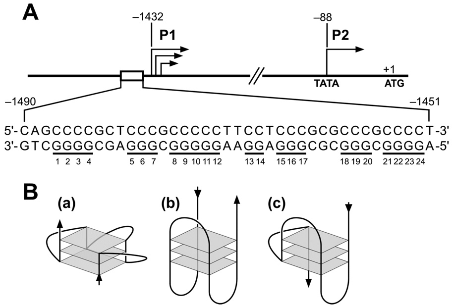
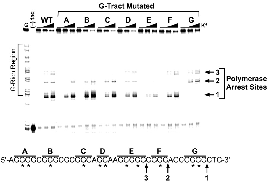
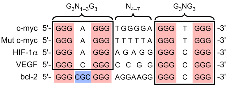
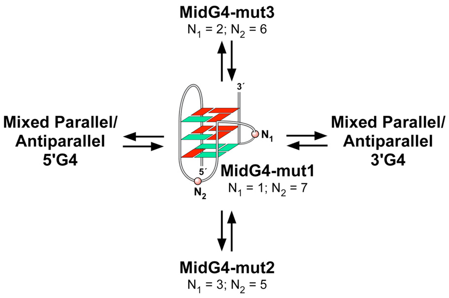
The human bcl-2 gene contains a GC-rich region upstream of the P1 promoter that has been shown to be critically involved in the regulation of bcl-2 gene expression. We have demonstrated that the guanine-rich strand of the DNA in this region can form any one of three distinct intramolecular G-quadruplex structures. Mutation and deletion analysis permitted isolation and identification of three overlapping DNA sequences within this element that formed the three individual G-quadruplexes. Each of these was characterized using nondenaturing gel analysis, DMS footprinting, and circular dichroism. The central G-quadruplex, which is the most stable, forms a mixed parallel/antiparallel structure consisting of three tetrads connected by loops of one, seven, and three bases. Three different G-quadruplex-interactive agents were found to further stabilize these structures, with individual selectivity toward one or more of these G-quadruplexes. Collectively, these results suggest that the multiple G-quadruplexes identified in the promoter region of the bcl-2 gene are likely to play a similar role to the G-quadruplexes in the c-myc promoter in that their formation could serve to modulate gene transcription. Last, we demonstrate that the complexity of the G-quadruplexes in the bcl-2 promoter extends beyond the ability to form any one of three separate G-quadruplexes to each having the capacity to form either three or six different loop isomers. These results are discussed in relation to the biological significance of this G-quadruplex-forming element in modulation of bcl-2 gene expression and the inherent complexity of the system where different G-quadruplexes and loop isomers are possible.


Figures

Figure 1
Figure 1
(A) Promoter structure of the human bcl-2 gene; shown in inset is the 39-mer sequence of the purine-rich strand upstream of the P1 promoter. Runs of guanines are underlined and numbered. (B) Examples of intramolecular G-quadruplex structures: (a) parallel, (b) antiparallel, (c) mixed parallel/antiparallel.
Figure 2
Figure 2
Structures of the G-quadruplex-interactive compounds TMPyP4, telomestatin, and Se2SAP, and the control compound TMPyP2.
Figure 3
Figure 3
Determination of the ability of the Pu39WT sequence (Table 1) to form multiple G-quadruplex structures. (A) DNA polymerase stop assay showing three K+-dependent DNA polymerase arrests. Lane 1 shows a guanine sequencing reaction, lane 2 is a negative control containing no Taq DNA polymerase, and lanes 3–6 contain 0, 25, 50, and 100 mM KCl, respectively. Arrows point to the positions of the full-length product of DNA synthesis, the DNA polymerase arrest sites (1, 2, and 3), and the free primer. (B) Concentration-dependent inhibition of Taq polymerase DNA synthesis by stabilization of the bcl-2 G-quadruplex structures with TMPyP4 (0.5, 1, 2.5, 5 µM) compared to TMPyP2 (0.5, 1, 2.5, 5 µM). All lanes contain 20 mM KCl. (C) Nondenaturing gel analysis of Pu39WT sequence preincubated in the absence or presence of 100 mM KCl. (D) DMS footprinting of band 1 (lane 2) and band 2 (lane 3) from (C). Lane 1 shows a purine sequencing reaction. The protected guanines from DMS are indicated with filled circles, partial protection with half-filled circles, no protection with open circles, and hypersensitive guanines with arrowheads. (E) Comparative CD spectra of three known G-quadruplex-forming sequences with the unknown bcl-2 promoter Pu39WT sequence. Line colors: green = c-myc promoter sequence (parallel G-quadruplex in 100 mM KCl); blue = thrombin binding aptamer (TBA) sequence (antiparallel G-quadruplex in 100 mM KCl); red = Tetrahymena telomeric repeat sequence (mixed parallel/antiparallel G-quadruplex in 100 NaCl); black = Tetrahymena telomeric repeat sequence (100 mM KCl); orange = bcl-2 promoter Pu39WT sequence (100 mM KCl). All CD data were obtained with a 5 µM strand concentration at 25 °C.
Figure 4
Figure 4
DNA polymerase stop assay to analyze the effect on stability of the three individual G-quadruplex structures formed by the Pu39WT sequence by knocking-out each G-tract with mutations. In the sequence below the gel, letters (A–G) indicate which G-tract contains the mutation(s) and asterisks indicate G-to-T mutations. The three DNA polymerase arrest sites (1, 2, and 3) are identified with arrows. KCl concentrations are 0, 50, and 100 mM.
Figure 5
Figure 5
Characterization of the three individual G-quadruplex-forming sequences. (A′–C′) DNA polymerase stop assays utilizing the 5′G4, MidG4, and 3′G4 sequences (see Table 1). Arrows point to the positions of the K+-dependent DNA polymerase arrest sites (G8, G5, and G1). KCl concentrations are 0, 25, 50, and 100 mM. (A″–C″) DMS footprinting and graphical representation of autoradiograms of isolated bands from the nondenaturing gels shown in the inset. Nondenaturing gel analysis of 5′G4, MidG4, and 3′G4 sequences preincubated under the conditions specified in the figure are shown in panel insets. The guanines protected from DMS are indicated with filled circles, partial protection with half-filled circles, no protection with open circles, and hypersensitive guanines with arrowheads. (D) The top panel shows comparative CD spectra of the bcl-2 promoter full-length and truncated sequences in the presence of 100 mM KCl. Line colors: orange = Pu39WT; red = 5′G4; blue = MidG4; black = 3′G4. The bottom panel compares the Tetrahymena telomeric repeat sequence with the bcl-2 MidG4 sequence. Line colors: red = Tetrahymena telomeric repeat sequence (mixed parallel-antiparallel G-quadruplex in 100 NaCl); black = Tetrahymena telomeric repeat sequence (100 mM KCl); blue = MidG4 (100 mM KCl); orange = bcl-2 promoter Pu39WT sequence (100 mM KCl). All CD data were obtained with a 5 µM strand concentration at 25° C.
Figure 6
Figure 6
DNA polymerase stop assay showing the difference in stability of the three possible loop isomers in the MidG4 sequence in comparison to the wild-type sequence. The wild-type MidG4 sequence is shown below the gel. MidG4-mut1, MidG4-mut2, and MidG4-mut3 contain dual G-to-T mutations within G5 (see Table 1). KCl concentrations are 0, 25, 50, and 100 mM.
Figure 7
Figure 7
Stabilization of the three individual G-quadruplex structures formed by the 5′G4, MidG4, and 3′G4 sequences with the addition of increasing concentrations of TMPyP2 (0.1, 0.25, 0.5, 1, and 2.5 µM), TMPyP4 (0.1, 0.25, 0.5, 1, and 2.5 µM), Se2SAP (0.05, 0.1, 0.25, 0.5, and 1 µM), and telomestatin (0.05, 0.1, 0.25, 0.5, and 1 µM) in the presence of 20 mM KCl. Quantification of autoradiograms is shown in right panels.
Figure 8
Figure 8
Comparison of truncated G-quadruplex-forming sequences within selected gene promoters.
Figure 9
Figure 9
Proposed equilibrium between the multiple G-quadruplex structures formed within the bcl-2 promoter region (red = anti conformation; green = syn conformation). The G-quadruplex folding pattern is as determined by an NMR study.

Similar articles

Cited by

References

    1. Chao DT, Korsmeyer SJ. Annu. Rev. Immunol. 1998;16:395–419. - PubMed
    1. Adams JM, Cory S. Science. 1998;281:1322–1326. - PubMed
    1. Yunis JJ. Science. 1983;221:227–236. - PubMed
    1. Cleary ML, Smith SD, Sklar J. Cell. 1986;47:19–28. - PubMed
    1. Akagi T, Kondo E, Yoshino T. Leuk. Lymphoma. 1994;13:81–87. - PubMed

Publication types