. 2006 Jun;154(3):269-79.
doi: 10.1016/j.jsb.2006.02.011. Epub 2006 Mar 24.

The three-dimensional structure of complex I from Yarrowia lipolytica: a highly dynamic enzyme

Affiliations

The three-dimensional structure of complex I from Yarrowia lipolytica: a highly dynamic enzyme

M Radermacher et al. J Struct Biol. 2006 Jun.

Abstract

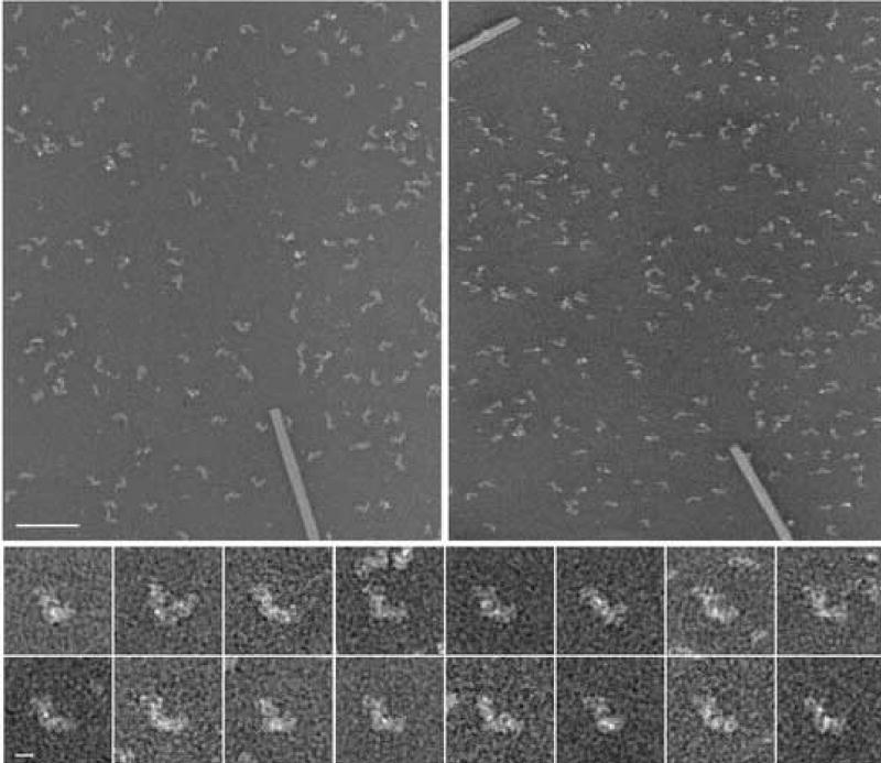
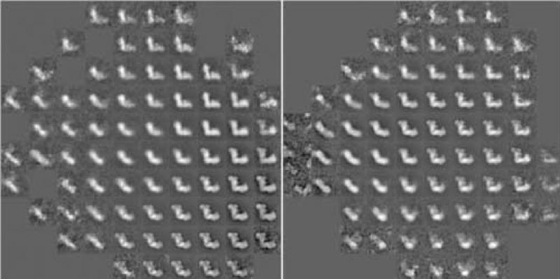
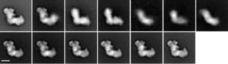
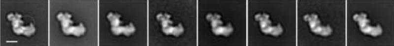
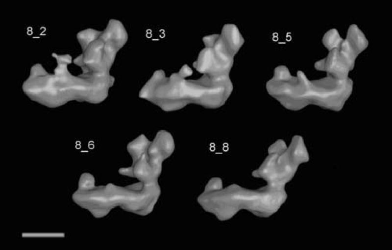
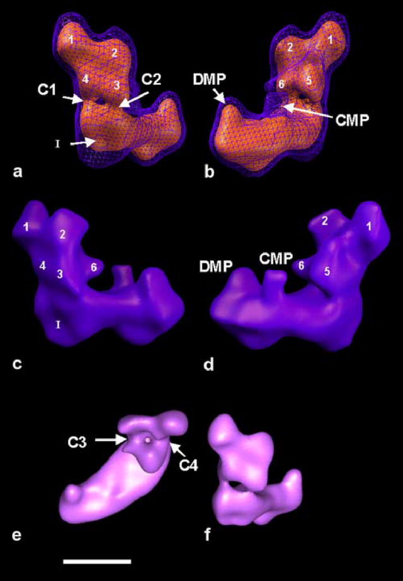
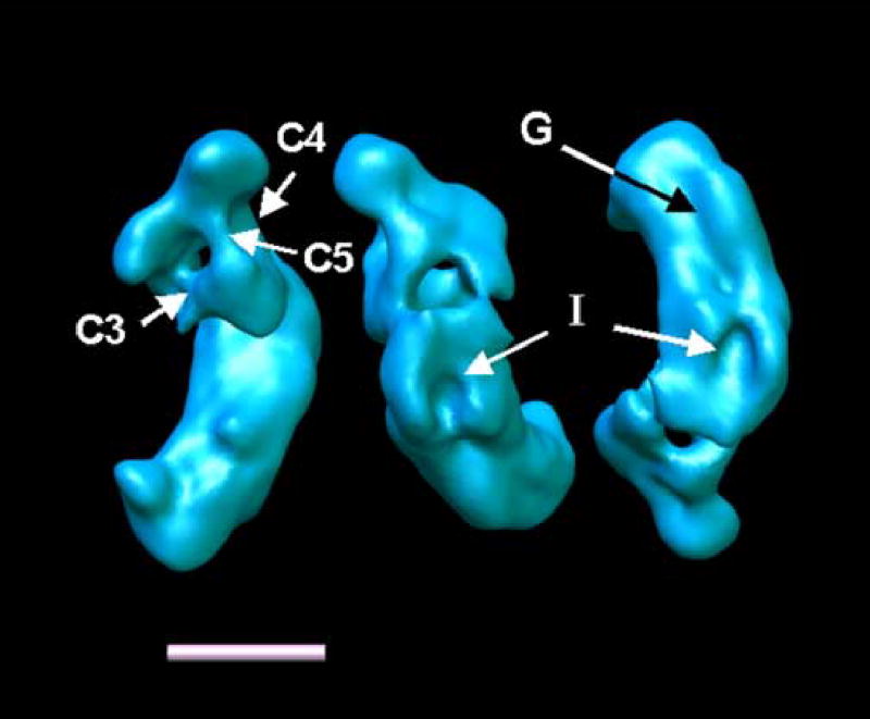
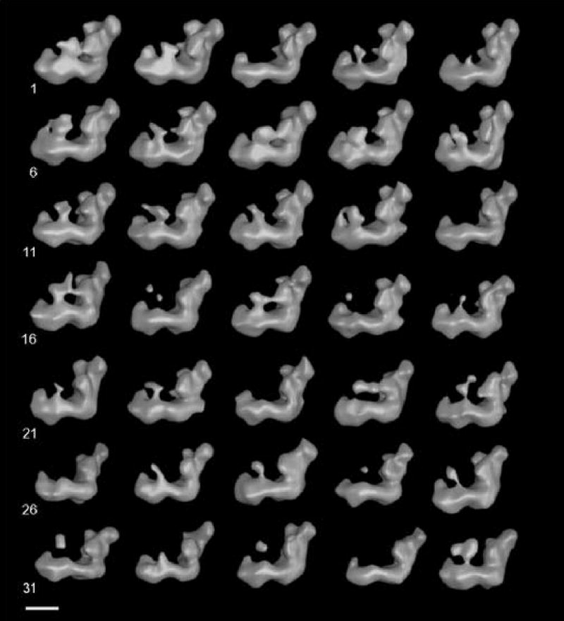
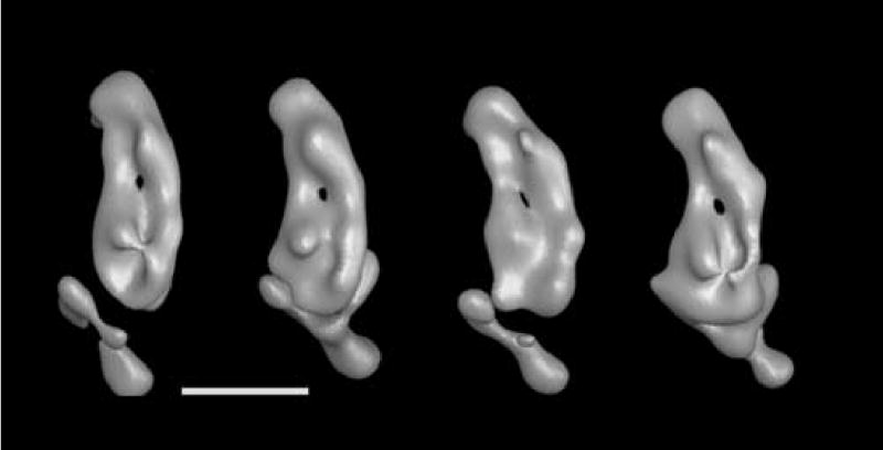
The structure of complex I from Yarrowia lipolytica was determined by three-dimensional electron microscopy. A random conical data set was collected from deep stain embedded particles. More than 14000 image pairs were analyzed. Through extensive classification combined with three-dimensional reconstruction, it was possible for the first time to show a much more detailed substructure of the complex. The peripheral arm is subdivided in at least six domains. The membrane arm shows two major protrusions on its matrix facing side and exhibits a channel like feature on the side facing the cytoplasm. Structures resembling a tether connecting the subunits near the catalytic center with the protrusions of the membrane arm provide a second connection between matrix and membrane domain.


Figures

Figure 1
Figure 1
Tilt pair of complex I from Y. lipolytica, prepared in deep stain (PTA). For a reconstruction from a random conical data set, pairs of micrographs of each specimen area are recorded, the first with a high tilt angle and the second without tilt. The particles from 0° micrographs are used for alignments and classification and three-dimensional reconstructions are calculated from the corresponding particles extracted from the tilt micrographs. The right image was recorded at a tilt angle of 55°, the left image at 0°tilt. TMV was used for the calibration of the magnification. Tilt axis horizontal, Scale bar 100 nm. Bottom: Series of aligned 0° images, Scale bar 10 nm.
Figure 2
Figure 2
Correspondence analysis was carried out with a total of 12 factors. Shown are visual representation of factor maps 1 versus 2 (left) and 3 versus 4 (right) of the complete data set. The two maps present an overview over the major variations found in the data set.
Figure 3
Figure 3
Class-averages of the complete data set calculated from the run of correspondence analysis shown in fig. 2. Classes are shown in the order of their class number. Class memberships: (class number followed by number of particles): (1) 1552, (2) 686, (3) 580, (4) 920, (5) 131.00, (6) 431, (7) 1092, (8) 1352, (9) 1580, (10) 1672, (11) 1975, (12) 2328, (13) 427.
Figure 4
Figure 4
The eight classes after separate multireference alignment, correspondence analysis and classification. Class memberships: (1) 507, (2) 1921, (3) 1960, (4) 112, (5) 1674, (6) 2538, (7) 172, (8) 2002. Scale bar 10 nm. The circle in the left most image indicates the mask used for the classification into 35 classes shown in figure 8.
Figure 5
Figure 5
Surface representation of the reconstruction from the five largest classes of the “flip”-view analysis. The classes shown are top left to bottom right: 8_2, 8_3, 8_5, 8_6, 8_8.
Figure 6
Figure 6
The reconstruction from the full “flip-view” set. Indicated are the common features found in the majority of reconstructions. Distinguishable domains are numbered. DMP distal membrane arm protuberance, CMP central Membrane arm protuberance. C1-C4 connections. a-d) show the particle with the membrane arm approximately parallel to the horizontal. a) shows the particle rotated with the membrane arm pointing into the image, so that the back-side of the matrix arm is best visible. b) is rotated around the z-axis by approximately 180° relative to a). c) and d) are side views of the particle, related by ~180° and approximately perpendicular to their preferred orientation on the specimen support. e) shows a top view of the particle, as seen from the side of the mitochondrial matrix. f) for comparison the same surface level as seen from the back of the matrix arm. a) and b) show a combination of two threshold levels, the blue mesh includes a calculated volume of ~1.3 MDa and the red surface ~670 kDa. At limited resolution, these measurements are usually up to 30% larger than the actual enclosed protein volume. In b) and c) the same surface threshold was used as for the blue mesh in a) and b). Scale bar 10 nm.
Figure 7
Figure 7
Reconstruction 8_2 at a contour level that reveals the connections between the domains in the peripheral arm. C5 is a connection that can be observed in many reconstructions with varying density. I Indentation (same as in Fig. 8). G groove along the cytoplasmic side of the membrane arm. Shown are: left: a top view of complex I, center: a view of the bottom of the back of the matrix arm, right: a view of the cytoplasmic side of the membrane arm.
Figure 8
Figure 8
The 35 reconstructions. Clearly visible is the variable density connected to the central membrane arm protuberance. Scale bar 10 nm
Figure 9
Figure 9
From left to right: Volumes 35_2, 35_13, 35_15 and 35_20 from figure 8 shown here at a higher threshold value that reveals the groove in the cytoplasmic side of the membrane arm and the possible channel through the membrane arm. All views are from the cytoplasmic side of the membrane arm.

References

    1. Abdrakhmanova A, Zickermann V, Bostina M, Radermacher M, Schagger H, Kerscher S, Brandt U. Subunit composition of mitochondrial complex I from the yeast Yarrowia lipolytica. Biochim Biophys Acta. 2004;1658:148–156. - PubMed
    1. BBA . Special Issue. Biochimica et Biophysica Acta. 1998;1364:89–296. - PubMed
    1. Belogrudov G, Hatefi Y. Catalytic sector of complex I (NADH:ubiquinone oxidoreductase): subunit stoichiometry and substrate-induced conformation changes. Biochemistry. 1994;33:4571–4576. - PubMed
    1. Böttcher B, Scheide D, Hesterberg M, Nagel-Steger L, Friedrich T. A novel, enzymatically active conformation of the Escherichia coli ADH:ubiquinone oxidoreductase (complex I) Journal of Biological Chemistry. 2002;277:17970–17977. - PubMed
    1. Brandt U. Energy converting NADH:quinone oxidoreductase (Complex I) Ann Rev Biochem. 2006;75 in press. - PubMed

Publication types

MeSH terms