. 2006 May;188(9):3199-207.
doi: 10.1128/JB.188.9.3199-3207.2006.

Mutational analysis of the Escherichia coli melR gene suggests a two-state concerted model to explain transcriptional activation and repression in the melibiose operon

Affiliations

Mutational analysis of the Escherichia coli melR gene suggests a two-state concerted model to explain transcriptional activation and repression in the melibiose operon

Christina Kahramanoglou et al. J Bacteriol. 2006 May.

Abstract

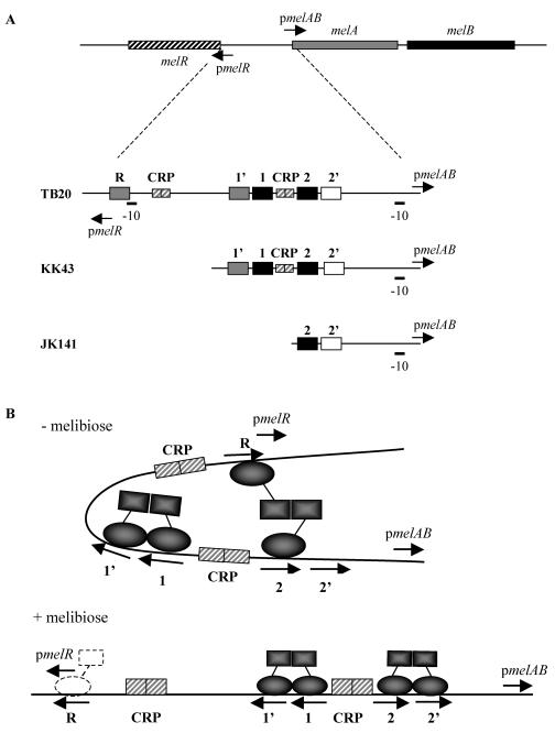
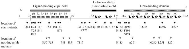
Transcription of the Escherichia coli melAB operon is regulated by the MelR protein, an AraC family member whose activity is modulated by the binding of melibiose. In the absence of melibiose, MelR is unable to activate the melAB promoter but autoregulates its own expression by repressing the melR promoter. Melibiose triggers MelR-dependent activation of the melAB promoter and relieves MelR-dependent repression of the melR promoter. Twenty-nine single amino acid substitutions in MelR that result in partial melibiose-independent activation of the melAB promoter have been identified. Combinations of different substitutions result in almost complete melibiose-independent activation of the melAB promoter. MelR carrying each of the single substitutions is less able to repress the melR promoter, while MelR carrying some combinations of substitutions is completely unable to repress the melR promoter. These results argue that different conformational states of MelR are responsible for activation of the melAB promoter and repression of the melR promoter. Supporting evidence for this is provided by the isolation of substitutions in MelR that block melibiose-dependent activation of the melAB promoter while not changing melibiose-independent repression of the melR promoter. Additional experiments with a bacterial two-hybrid system suggest that interactions between MelR subunits differ according to the two conformational states.


Figures

FIG. 1.
FIG. 1.
Organization of the E. coli melibiose operon regulatory region. (A) A not-to-scale illustration of the organization of the melR, melA, and melB genes, with the locations and orientation of pmelR and pmelAB. In the lower part of the figure, expanded views of the TB20, KK43, and JK141 fragments are shown, with the locations of the pmelAB and pmelR −10 elements and the different DNA sites for CRP (small hatched boxes) and MelR (larger boxes shaded according to binding hierarchy in the absence of melibiose). In this work, the TB20 fragment was cloned with EcoRI and HindIII linkers upstream and downstream of pmelR, respectively, into pRW50 to give a pmelR::lac fusion. The KK43 and JK141 fragments were cloned with EcoRI and HindIII linkers upstream and downstream of pmelAB, respectively, into pRW50 to give a pmelAB::lac fusion. (B) Interactions of MelR with the different sites in the absence and presence of melibiose as proposed by Wade et al. (30). In the absence of melibiose, MelR is unable to occupy site 2′, and an interaction between MelR bound at site 2 and site R causes strong repression of pmelR. In the presence of melibiose, MelR occupies site 2′, the interaction between site 2 and site R is broken, and the strong repression of pmelR is relieved. Weaker repression is due to residual binding of MelR to site R (dotted outline).
FIG. 2.
FIG. 2.
Domain organization of the E. coli MelR protein. The 302 amino acids of MelR are illustrated as a horizontal line, annotated with different structural features. The locations of a ligand-binding cupin fold, consisting of eight β-sheet elements (5, 6) and a helix-loop-helix dimerization motif, deduced from similarities with AraC and published AraC structures (26, 27), are shown. The location of the DNA-binding domain, consisting of seven α-helix elements and deduced from similarities with MarA and the published MarA structure (20), is shown. The lower part of the figure shows the locations of different substitutions that confer the ability to partially activate pmelAB in the absence of melibiose (stars) and the locations of substitutions that interfere with melibiose-dependent activation (diamonds).

References

    1. Barne, K. A., J. A. Bown, S. J. Busby, and S. D. Minchin. 1997. Region 2.5 of the Escherichia coli RNA polymerase sigma70 subunit is responsible for recognition of the extended −10′ motif at promoters. EMBO J. 16:4034-4040. - PMC - PubMed
    1. Belyaeva, T. A., J. T. Wade, C. L. Webster, V. J. Howard, M. S. Thomas, E. I. Hyde, and S. J. Busby. 2000. Transcription activation at the Escherichia coli melAB promoter: the role of MelR and the cyclic AMP receptor protein. Mol. Microbiol. 36:211-222. - PubMed
    1. Bustos, S. A., and R. F. Schleif. 1993. Functional domains of the AraC protein. Proc. Natl. Acad. Sci. USA 90:5638-5642. - PMC - PubMed
    1. Datsenko, K. A., and B. L. Wanner. 2000. One-step inactivation of chromosomal genes in Escherichia coli K-12 using PCR products. Proc. Natl. Acad. Sci. USA 97:6640-6645. - PMC - PubMed
    1. Dunwell, J. M., A. Culham, C. E. Carter, C. R. Sosa-Aguirre, and P. W. Goodenough. 2001. Evolution of functional diversity in the cupin superfamily. Trends Biochem. Sci. 26:740-746. - PubMed

Publication types

LinkOut - more resources