. 2006 May;188(9):3273-9.
doi: 10.1128/JB.188.9.3273-3279.2006.

Mutation of waaC, encoding heptosyltransferase I in Campylobacter jejuni 81-176, affects the structure of both lipooligosaccharide and capsular carbohydrate

Affiliations

Mutation of waaC, encoding heptosyltransferase I in Campylobacter jejuni 81-176, affects the structure of both lipooligosaccharide and capsular carbohydrate

Margaret I Kanipes et al. J Bacteriol. 2006 May.

Abstract

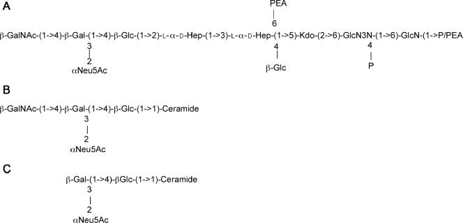
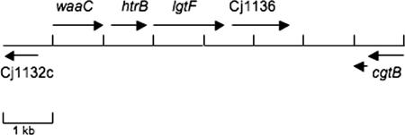
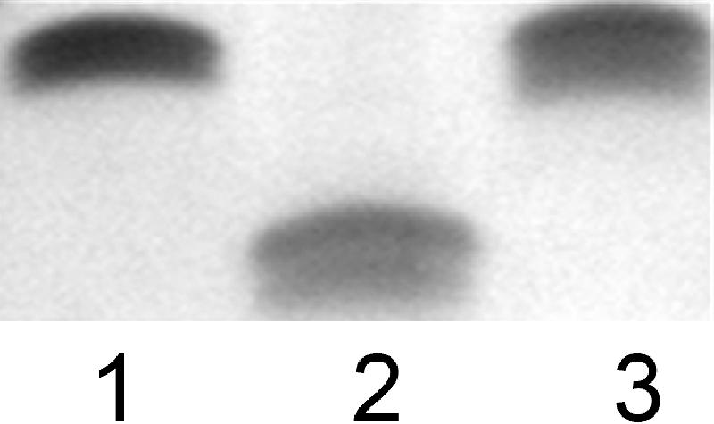
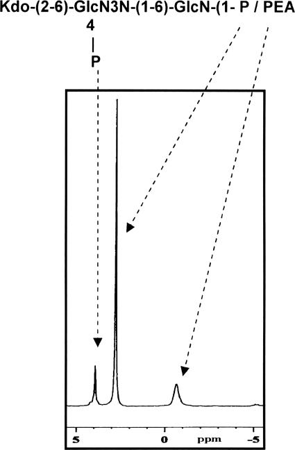
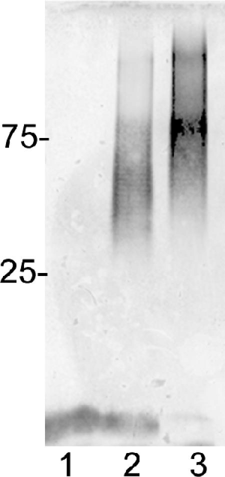
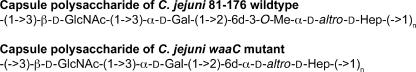
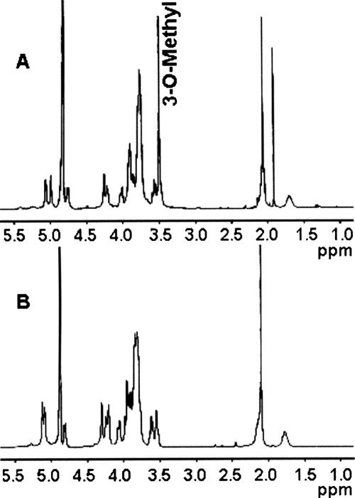
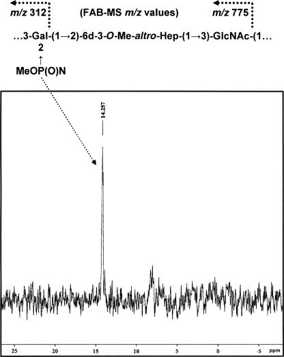
Campylobacter jejuni 81-176 lipooligosaccharide (LOS) is composed of two covalently linked domains: lipid A, a hydrophobic anchor, and a nonrepeating core oligosaccharide, consisting of an inner and outer core region. We report the isolation and characterization of the deepest rough C. jejuni 81-176 mutant by insertional mutagenesis into the waaC gene, encoding heptosyltransferase I that catalyzes the transfer of the first L-glycero-D-manno-heptose residue to 3-deoxy-D-manno-octulosonic residue (Kdo)-lipid A. Tricine gel electrophoresis, followed by silver staining, showed that site-specific mutation in the waaC gene resulted in the expression of a severely truncated LOS compared to wild-type strain 81-176. Gas-liquid chromatography-mass spectrometry and nuclear magnetic resonance spectroscopy showed that the waaC LOS species lacked all sugars distal to Kdo-lipid A. Parallel structural studies of the capsular polysaccharides of the wild-type strain 81-176 and waaC mutant revealed loss of the 3-O-methyl group in the waaC mutant. Complementation of the C. jejuni mutant by insertion of the wild-type C. jejuni waaC gene into a chromosomal locus resulted in LOS and capsular structures identical to those expressed in the parent strain. We also report here the presence of O-methyl phosphoramidate in wild-type strain 81-176 capsular polysaccharide.


Figures

FIG. 1.
FIG. 1.
(A) Core oligosaccharide of C. jejuni 81-176 (serogroups O:23/O:36) and the phosphorylated disaccharide backbone of lipid A (determined for strain CCUG 10936) (23). The structure of 81-176 LOS has been reported to vary between the structure shown in panel A, which mimics GM3 ganglioside (B), and a structure lacking the terminal GalNAc, which mimics GM2 ganglioside (C). This variation is due to slip strand mismatch repair of the cgtA gene, encoding a GalNAc transferase (12).
FIG. 2.
FIG. 2.
Schematic representation of the C. jejuni 81-176 chromosomal region encoding relevant LOS genes. The overall genomic organization is similar to that of class B loci described by Gilbert and colleagues and Parker et al. (7, 28). The ORF designated waaC showed 87% identity and 88% similarity to WaaC (Cj1133) from the C. jejuni genome strain NCTC11168. The cat transposon insertion that was used to generate the mutation was at 677 bp from the translational start of the 1,029-bp waaC gene. The second ORF encoded a predicted protein with 99% identity to HtrB, a lipid A biosynthesis lauroyl acyltransferase, from C. jejuni strain GB11. The third ORF encoded a protein that showed 88% identity and 94% similarity to a putative two-domain glycosyltransferase of C. jejuni NCTC11168 (Cj1135). The fourth ORF exhibited 75% identity and 86% similarity to a putative β-1,3-galactosyltransferase from C. jejuni NCTC1168 (Cj1136). The 81-176 homolog of cgtB was slip stranded out of frame as cloned. This gene encodes a galactosyltransferase that has been shown to undergo phase variation in another strain to convert GM2 to GM1 mimics (20). Between Cj1136 and the cgtB homolog, there was no ORF of >174 bp, but BLASTX analysis revealed homology to a β-1,4-GalNAc transferase, similar to what has been reported for other class B LOS loci (7, 28).
FIG. 3.
FIG. 3.
LOS core mobility. Proteinase K-digested whole-cell preparations were electrophoresed on 16% Tricine gels and silver stained. Lane 1, 81-176; lane 2, waaC mutant; and lane 3, complement of waaC mutant.
FIG. 4.
FIG. 4.
31P NMR spectrum of the truncated waaC LOS showing phosphorus resonances for ester-bound phosphate and for glycosidic-linked phosphate residues.
FIG. 5.
FIG. 5.
Immunoblot of C. jejuni waaC capsular material from a 12% SDS-PAGE gel. Lane 1, 81-176 kpsM mutant (2); lane 2, 81-176; and lane 3, 81-176 waaC mutant. The positions of protein markers in kDa are shown at the left. The LOS cores of the 81-176 kpsM mutant and the wild type are visible at the bottom of the immunoblot; the core of the waaC mutant is not visible.
FIG. 6.
FIG. 6.
The reported CPS structures of C. jejuni O:23/O:36 and 81-176 strains (32) and the 81-176 waaC mutant. Abbreviations: 6d-3-O-Me-altro-Hep, 6-deoxy-3-O-methyl-altro-heptose; 6d-altro-Hep, 6-deoxy-altro-heptose.
FIG. 7.
FIG. 7.
NMR analyses. (A) 1H NMR of C. jejuni 81-176 wild-type CPS (1% acetic acid treated) showing (i) the anomeric resonances at δ 5.06 for α-Gal, δ 4.99 for 3-O-Me-6-deoxy-α-altro-Hep, and δ 4.75 for β-GlcNAc, (ii) the methyl signal at δ 3.50 for 3-O-Me-6-deoxy-α-altro-Hep, (iii) the methyl resonance at δ 2.09 for 3-O-Me-6-deoxy-α-altro-Hep, and (iv) the deoxy resonance at δ 1.70 for 3-O-Me-6-deoxy-α-altro-Hep. (B) 1H NMR of C. jejuni 81-176 waaC CPS showing (i) the anomeric resonances at δ 5.12 for 6-deoxy-α-altro-Hep, δ 4.98 for α-Gal, and δ 4.75 for β-GlcNAc, (ii) the methyl signal at δ 2.09 for GlcNAc, and (iii) the deoxy resonance at δ 1.75 for GlcNAc. No 3-O-methyl signal (3-O-Me-6-deoxy-α-altro-Hep) was observed in the waaC CPS 1H NMR spectrum. Also of note, the α-anomeric signal of 6-deoxy-α-altro-Hep in the waaC CPS resonated at δ 5.12, whereas the α-anomeric signal of 3-O-Me-6-deoxy-α-altro-Hep in wild-type CPS resonated at δ 4.99. The peak at δ 1.9 in panel B represents contaminating sodium acetate. All the results described were assigned by 2D 1H-1H NMR spectroscopy (data not shown).
FIG. 8.
FIG. 8.
The 31P NMR spectrum of C. jejuni wild-type 81-176 CPS showed a resonance at 14.257, attributable to an O-methyl phosphoramidate component, and the FAB-MS primary ions of the methylated wild-type 81-176 CPS indicated that O-methyl phosphoramidate is attached to the Gal unit in the CPS.

References

    1. Bacon, D. J., R. A. Alm, D. H. Burr, L. Hu, D. J. Kopecko, C. P. Ewing, T. J. Trust, and P. Guerry. 2000. Involvement of a plasmid in virulence of Campylobacter jejuni 81-176. Infect. Immun. 68:4384-4390. - PMC - PubMed
    1. Bacon, D. J., C. M. Szymanski, D. H. Burr, R. P. Silver, R. A. Alm, and P. Guerry. 2001. A phase-variable capsule is involved in virulence of Campylobacter jejuni 81-176. Mol. Microbiol. 40:769-777. - PubMed
    1. Black, R. E., M. M. Levine, M. L. Clements, T. P. Hughes, and M. J. Blaser. 1988. Experimental Campylobacter jejuni infection in humans. J. Infect. Dis. 157:472-479. - PubMed
    1. Ciucanu, I., and F. Kerek. 1984. A simple and rapid method for the permethylation of carbohydrates. Carbohydr. Res. 131:209-217.
    1. Dubois, M., K. Giles, J. Hamilton, P. Rebers, and F. Smith. 1956. Colometric method for determination of sugars and related substances. Anal. Chem. 28:350-356.

Publication types

MeSH terms

Associated data

LinkOut - more resources