. 2006 May;188(9):3345-56.
doi: 10.1128/JB.188.9.3345-3356.2006.

Biochemical bases of type IV chromatic adaptation in marine Synechococcus spp

Affiliations

Biochemical bases of type IV chromatic adaptation in marine Synechococcus spp

Craig Everroad et al. J Bacteriol. 2006 May.

Abstract

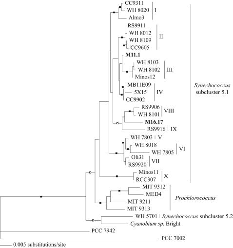
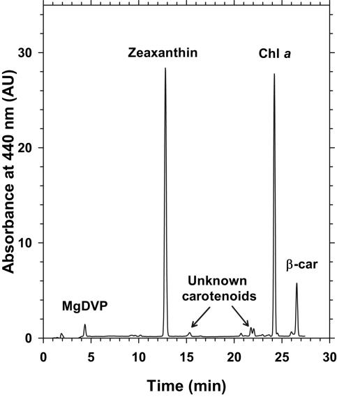
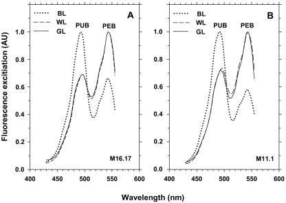
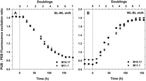
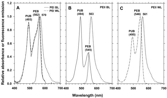
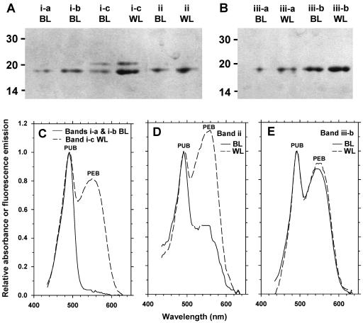
Chromatic adaptation (CA) in cyanobacteria has provided a model system for the study of the environmental control of photophysiology for several decades. All forms of CA that have been examined so far (types II and III) involve changes in the relative contents of phycoerythrin (PE) and/or phycocyanin when cells are shifted from red to green light and vice versa. However, the chromophore compositions of these polypeptides are not altered. Some marine Synechococcus species strains, which possess two PE forms (PEI and PEII), carry out another type of CA (type IV), occurring during shifts from blue to green or white light. Two chromatically adapting strains of marine Synechococcus recently isolated from the Gulf of Mexico were utilized to elucidate the mechanism of type IV CA. During this process, no change in the relative contents of PEI and PEII was observed. Instead, the ratio of the two chromophores bound to PEII, phycourobilin and phycoerythrobilin, is high under blue light and low under white light. Mass spectroscopy analyses of isolated PEII alpha- and beta-subunits show that there is a single PEII protein type under all light climates. The CA process seems to specifically affect the chromophorylation of the PEII (and possibly PEI) alpha chain. We propose a likely process for type IV CA, which involves the enzymatic activity of one or several phycobilin lyases and/or lyase-isomerases differentially controlled by the ambient light quality. Phylogenetic analyses based on the 16S rRNA gene confirm that type IV CA is not limited to a single clade of marine Synechococcus.


Figures

FIG. 1.
FIG. 1.
Neighbor-joining phylogeny inferred from partial 16S rRNA gene sequences rooted with Synechococcus strain PCC 7002 as the outgroup. Closed boxes indicate bootstrap values of >70 for both neighbor-joining and maximum parsimony analyses and a Bayesian posterior probability of >0.85. Open boxes designate support as described above but only for neighbor-joining and Bayesian analyses. Roman numerals indicate the 10 clades of Synechococcus subcluster 5.1 identified by Fuller and coworkers (8). Strains M11.1 and M16.17 are in bold.
FIG. 2.
FIG. 2.
HPLC pigment analysis (absorbance at 440 nm) of Synechococcus sp. strain M16.17 acclimated to blue light. β-car, β-carotene; Chl a, chlorophyll a; MgDVP, Mg-divinyl-phaeoporphyrin a5.
FIG. 3.
FIG. 3.
Fluorescence excitation spectra (with emission at 580 nm) of Synechococcus species strains M16.17 (A) and M11.1 (B) acclimated to white light (WL), green light (GL), and blue light (BL).
FIG. 4.
FIG. 4.
Acclimation kinetics of the Ex495 nm/Ex545 nm ratio after a shift (at time zero) from blue to white light (BL-WL) (A) and white to blue light (WL-BL) (B) in Synechococcus species strains M16.17 and M11.1.
FIG. 5.
FIG. 5.
Acclimation kinetics of flow cytometric parameters for Synechococcus species strains M16.17 (white symbols) and M11.1 (black symbols) after a shift (at time zero) from blue to white light (BL-WL) (A) and white to blue light (WL-BL) (B). FALS (circles) is a proxy for cell size, and orange fluorescence (squares) is a proxy for the PUB/PEB ratio.
FIG. 6.
FIG. 6.
Absorption spectra of intact PBSs from Synechococcus sp. strain M16.17 grown in blue light (BL) and white light (WL).
FIG. 7.
FIG. 7.
Coomassie blue-stained (left) and UV-visualized (right) LiDS-PAGE gels showing the linker composition of intact PBSs from Synechococcus species strains M11.1 and M16.17 grown in blue (BL) and white light (WL). The corresponding profile obtained for the chromatically nonadapting Synechococcus sp. strain WH 8102, in which all the linkers have been firmly identified (33), are shown for comparison. Note that the latter profile differs from the previously published one by an artifactual doubling of the MpeE band at ca. 38 kDa. Double arrows indicate the positions of LCM and LCM′ linker polypeptides (both of which fluoresce blue under UV light), and the single arrow indicates the likely position of FNR in each strain. Minor bands located between the FNR and MpeD in M11.1 are likely proteolytic fragments of MpeD.
FIG. 8.
FIG. 8.
Different purification steps of phycobilisome components from Synechococcus sp. strain M16.17 cells grown in blue light (BL) and white light (WL). Filtered whole cells (A); dissociation of soluble proteins on continuous sucrose gradient showing the different fractions 1, 2, and 3 (B); native IEF gel of soluble proteins showing separated PBP complexes, including two PEII fractions (a and b) (C); and denaturing IEF showing separated PBP polypeptides (D). See the text for details.
FIG. 9.
FIG. 9.
Optical properties of Synechococcus sp. strain M16.17 phycoerythrin complexes. PEI absorption spectra in blue light (BL; continuous line) and white light (WL; dashed line) and corresponding fluorescence emission spectra (identical for both light qualities; dotted line) (A); PEII absorption and fluorescence emission spectra in blue light (B) and white light (C). Note that profiles obtained for the PEII a and PEII b bands (Fig. 8C) were identical.
FIG. 10.
FIG. 10.
LiDS-PAGE (A and B) and absorption spectra (C, D, and E) of the main bands from the denaturing IEF gel shown in Fig. 8D, obtained from Synechococcus sp. strain M16.17 cells grown in blue light (BL) and white light (WL). The absorption spectra of bands i-a and i-b (present only in BL) were identical and very different from that of band i-c (C). The LiDS-PAGE analysis of band i-c (A) revealed that it was made up of two proteins, a minor (∼20-kDa) protein, CpeB, and a major (∼18-kDa) protein, MpeA, also found (pure) in bands i-a and i-b. Bands iii-a and iii-b contained a sole ∼19-kDa protein, MpeB (B).

References

    1. Alberte, R. S., A. M. Wood, T. A. Kursar, and R. R. L. Guillard. 1984. Novel phycoerythrins in marine Synechococcus spp. Plant Physiol. 75:732-739. - PMC - PubMed
    1. Anderson, L. K., M. C. Rayner, and F. A. Eiserling. 1984. Ultra-violet mutagenesis of Synechocystis sp. 6701: mutations in chromatic adaptation and phycobilisome assembly. Arch. Microbiol. 138:237-243.
    1. Brahamsha, B. 1996. A genetic manipulation system for oceanic cyanobacteria of the genus Synechococcus. Appl. Environ. Microbiol. 62:1747-1751. - PMC - PubMed
    1. Campbell, L., and R. Iturriaga. 1988. Identification of Synechococcus spp. in the Sargasso Sea by immunofluorescence and fluorescence excitation spectroscopy performed on individual cells. Limnol. Oceanogr. 33:1196-1201.
    1. Conley, P. B., P. G. Lemaux, and A. Grossman. 1988. Molecular characterization and evolution of sequences encoding light-harvesting components in the chromatically adapting cyanobacterium Fremyella diplosiphon. J. Mol. Biol. 199:447-465. - PubMed

Publication types

Associated data

LinkOut - more resources