. 2006 May;188(9):3391-401.
doi: 10.1128/JB.188.9.3391-3401.2006.

Structure of the functional form of the mosquito larvicidal Cry4Aa toxin from Bacillus thuringiensis at a 2.8-angstrom resolution

Affiliations

Structure of the functional form of the mosquito larvicidal Cry4Aa toxin from Bacillus thuringiensis at a 2.8-angstrom resolution

Panadda Boonserm et al. J Bacteriol. 2006 May.

Abstract

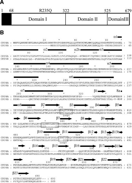
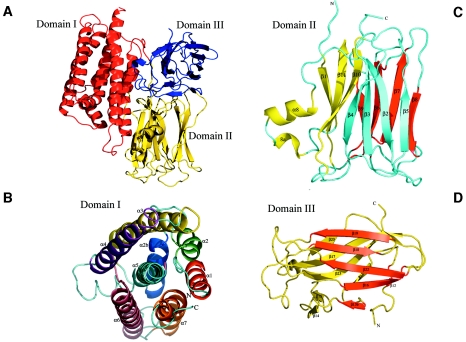
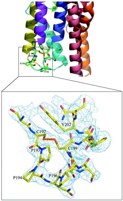
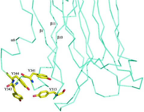
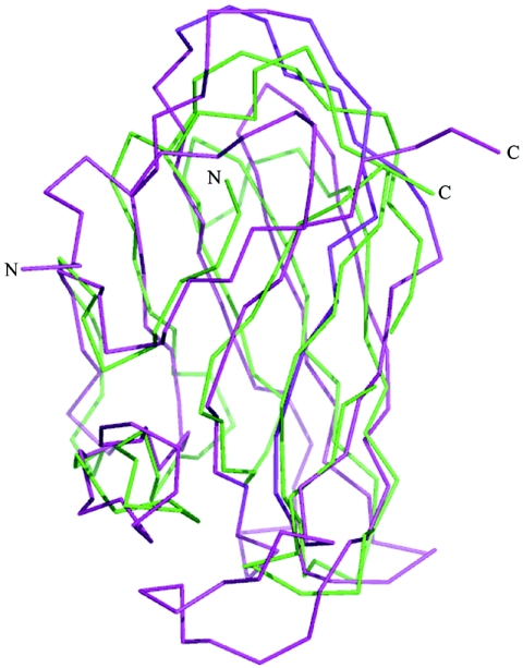
The Cry4Aa delta-endotoxin from Bacillus thuringiensis is toxic to larvae of Culex, Anopheles, and Aedes mosquitoes, which are vectors of important human tropical diseases. With the objective of designing modified toxins with improved potency that could be used as biopesticides, we determined the structure of this toxin in its functional form at a resolution of 2.8 angstroms. Like other Cry delta-endotoxins, the activated Cry4Aa toxin consists of three globular domains, a seven-alpha-helix bundle responsible for pore formation (domain I) and the following two other domains having structural similarities with carbohydrate binding proteins: a beta-prism (domain II) and a plant lectin-like beta-sandwich (domain III). We also studied the effect on toxicity of amino acid substitutions and deletions in three loops located at the surface of the putative receptor binding domain II of Cry4Aa. Our results indicate that one loop is an important determinant of toxicity, presumably through attachment of Cry4Aa to the surface of mosquito cells. The availability of the Cry4Aa structure should guide further investigations aimed at the molecular basis of the target specificity and membrane insertion of Cry endotoxins.


Figures

FIG. 1.
FIG. 1.
(A) Schematic representation of the three domains present in the mature Cry4Aa toxin. Amino acids 68 to 679 are visible in the final electron density map. The R235Q mutation, which makes the toxin resistant to further proteolysis, is also indicated. (B) Alignment of the amino acid sequences of the homologous dipteran-specific toxins Cry4Aa and Cry4Ba. Secondary structure elements of Cry4Aa, as determined in this work (using the Pymol program [16]), are indicated above the sequence; α helices are indicated by rectangles, and β strands are indicated by arrows. 8a is a short 310 helical segment. Residue numbers of both toxins are indicated.
FIG. 2.
FIG. 2.
Ribbon representations of the Cry4Aa fold. (A) Overall view of the Cry4Aa toxin, which is composed of three domains. Pore-forming domain I is red, putative receptor binding domain II is yellow, and C-terminal domain III is blue. The secondary structure elements of each of the three domains are labeled in panels B to D. Each individual β-sheet is a different color. See Fig. 1B.
FIG. 3.
FIG. 3.
Surface representation of the electrostatic potential of pore-forming domain I of Cry4Aa (calculated using Pymol [16]). Positive electrostatic potentials are blue, and negative electrostatic potentials are red. The locations of solvent-exposed α-helices are indicated. The solvent-accessible surfaces of α-helices α1, α6, and α7 (A) show relatively higher potential and clear charge separations (including a “basic strip”) than α-helices α3 and α4 (B). Panel B is a view with domain I rotated 180° along a vertical axis; this has implications for the way that domain I approaches the target cell membrane (see text).
FIG. 4.
FIG. 4.
“Open book” representation of the interfaces between domains I and II of Cry4Aa and Cry4Ba. The views were obtained by rotating each of the interacting domains approximately 90° around a vertical axis. A hydrophobic patch is visible at the interface between domains I and II. Amino acids L326, I330, Y331, V333, L334, and F336 in Cry4Aa and amino acids F287, I291, Y292, A294, L295, and V296 in Cry4Ba, which are present in this hydrophobic patch, are labeled.
FIG. 5.
FIG. 5.
View of the α4-α5 loop in domain I of Cry4Aa. This segment is thought to play an important role in membrane insertion (see text). At the bottom is a close-up of the region containing the α4-α5 loop indicated by the rectangle. The final electron density map is displayed at a contour of 1σ. There is a disulfide bridge between Cys-192 and Cys-199.
FIG. 6.
FIG. 6.
View of the exposed cluster of aromatic amino acids (indicated by sticks) at the surface of domain II of Cry4Aa (only a partial view of the α-carbon chain of domain II is shown). This cluster is proposed to function as a carbohydrate binding site (see text). Secondary structure elements are indicated.
FIG. 7.
FIG. 7.
Superposition of C-terminal domain III of Cry4Aa with xylanase from C. thermocellum, highlighting the common fold. Only the α-carbon traces are shown (magenta for Cry4Aa and green for xylanase).
FIG. 8.
FIG. 8.
Comparison of the structures adopted by the three surface-exposed loops of Cry4Aa and Cry4Ba at the extremity of domain II. Domain II of Cry4Aa is cyan, and domain II of Cry4Ba is yellow. The locations of loops 1, 2, and 3 on domain II are indicated, and close-ups highlight the structural differences in these putative receptor binding regions. With the possible exception of loop 1 of Cry4Ba, which makes several crystalline intermolecular contacts (7), the conformations of the other loops are likely to be determined by intramolecular contacts, not by crystal packing interactions.

References

    1. Abdullah, M. A., O. Alzate, M. Mohammad, R. J. McNall, M. J. Adang, and D. H. Dean. 2003. Introduction of Culex toxicity into Bacillus thuringiensis Cry4Ba by protein engineering. Appl. Environ. Microbiol. 69:5343-5353. - PMC - PubMed
    1. Angsuthanasombat, C., N. Crickmore, and D. J. Ellar. 1992. Comparison of Bacillus thuringiensis subsp. israelensis CryIVA and CryIVB cloned toxins reveals synergism in vivo. FEMS Microbiol. Lett. 73:63-68. - PubMed
    1. Angsuthanasombat, C., N. Crickmore, and D. J. Ellar. 1993. Effects on toxicity of eliminating a cleavage site in a predicted interhelical loop in Bacillus thuringiensis CryIVB delta-endotoxin. FEMS Microbiol. Lett. 111:255-261. - PubMed
    1. Aronson, A. I., C. Geng, and L. Wu. 1999. Aggregation of Bacillus thuringiensis Cry1A toxins upon binding to target insect larval midgut vesicles. Appl. Environ. Microbiol. 65:2503-2507. - PMC - PubMed
    1. Ballester, V. V., F. Granero, R. A. de Maagd, D. Bosch, J. L. Mensua, and J. Ferre. 1999. Role of Bacillus thuringiensis toxin domains in toxicity and receptor binding in the diamondback moth. Appl. Environ. Microbiol. 65:1900-1903. - PMC - PubMed

Publication types

MeSH terms

LinkOut - more resources