Clinical Trial
. 2006 Aug 15;108(4):1267-79.
doi: 10.1182/blood-2005-10-007252. Epub 2006 Apr 18.

Genetics of HUS: the impact of MCP, CFH, and IF mutations on clinical presentation, response to treatment, and outcome

Affiliations
Clinical Trial

Genetics of HUS: the impact of MCP, CFH, and IF mutations on clinical presentation, response to treatment, and outcome

Jessica Caprioli et al. Blood. .

Abstract

Hemolytic uremic syndrome (HUS) is a thrombotic microangiopathy with manifestations of hemolytic anemia, thrombocytopenia, and renal impairment. Genetic studies have shown that mutations in complement regulatory proteins predispose to non-Shiga toxin-associated HUS (non-Stx-HUS). We undertook genetic analysis on membrane cofactor protein (MCP), complement factor H (CFH), and factor I (IF) in 156 patients with non-Stx-HUS. Fourteen, 11, and 5 new mutational events were found in MCP, CFH, and IF, respectively. Mutation frequencies were 12.8%, 30.1%, and 4.5% for MCP, CFH, and IF, respectively. MCP mutations resulted in either reduced protein expression or impaired C3b binding capability. MCP-mutated patients had a better prognosis than CFH-mutated and nonmutated patients. In MCP-mutated patients, plasma treatment did not impact the outcome significantly: remission was achieved in around 90% of both plasma-treated and plasma-untreated acute episodes. Kidney transplantation outcome was favorable in patients with MCP mutations, whereas the outcome was poor in patients with CFH and IF mutations due to disease recurrence. This study documents that the presentation, the response to therapy, and the outcome of the disease are influenced by the genotype. Hopefully this will translate into improved management and therapy of patients and will provide the way to design tailored treatments.


Figures

Figure 1.
Figure 1.
Modeling of IVS2 –2A>G mutation in MCP. (A) The pET (MoBiTec) exon traps cloning vectors containing wild-type or mutated genomic DNA from intron 1 (IVS1 –62) to intron 4 (IVS4 +70) of MCP. The vectors were transfected into 293T cells and the products were analyzed by RT-PCR and sequencing. Arrows 2 and 3 are the primers used for RT-PCR. Arrow 4 is the primer used for sequencing of cDNA product. MCP2, MCP3, and MCP4 indicate exons II, III, and IV of MCP. (B) Ethidium bromide–stained 1.5% agarose gel of RT-PCR products from splicing assays of transfected 293T cells. The wild-type minigene generated a product of 624 bp containing MCP exons II, III, and IV, whereas the IVS2 –2A>G mutant minigene produced a product of 521 bp lacking exon III. The wild-type sequence inserted in the reverse orientation gave a product (246 bp) that did not contain any spliced product. (C) Sequencing of cloned RT-PCR product. The RT-PCR products were subcloned into pCR2.1-TOPO and sequenced. The IVS2–2A>G mutation results in skipping of exon III. This alteration predicts a 34–amino acid loss (62-95del) in SCR2 followed by 3 amino acid changes (G96I +Y97I + Y98T) and a premature stop at L99.
Figure 2.
Figure 2.
Summary of MCP mutations in non-Stx–HUS patients from our registry. The corresponding number of mutational events is indicated in parentheses. SCR indicates short consensus repeat; STP, serine-threonine-proline–rich domain; TM, transmembrane domain; and CT, cytoplasmic tail.
Figure 3.
Figure 3.
Haplotype analysis on markers flanking MCP gene of family no. 099 and of patient S222 212, both of Sardinian origin, showing that a common allele carrying the mutation is present. The mutation is present in homozygosity in 2 affected siblings (F166 099 and F167 099) in family no. 099 from nonconsanguineous parents, both carrying the mutation in heterozygosity. Of note, the 2 siblings developed HUS very early in life (before 4 years of age) whereas their father (F168 099) developed HUS in adulthood and their mother is still healthy. Three other healthy family members carry the mutation in heterozygosity (marked with ·). Circles indicate females; squares, males; filled symbols, affected individuals; and open symbols, unaffected individuals.
Figure 4.
Figure 4.
Expression and functional studies on MCP mutants. (A) Flow cytometry analysis of MCP (CD46) expression in PBMCs from 5 mutation carriers (C1X ho: patient F166 099, homozygous for the IVS1 –1G>C mutation; C1Y he: unaffected healthy carrier of family no. 024, heterozygous for the 147G>A mutation, mother of patients F106 and F108; R25X/C1Y: patient F106 24, compound heterozygous for the 218C>T and 147G>A mutations; C65R he: patient S207 199, heterozygous for the 338T>C mutation; 238-242del* he: patient S045 169, heterozygous for the 858-872del 15bp+875C>T mutation) and from healthy controls (Control, n = 6). Percentages of CD46+ cells and median fluorescence intensity (MFI) are presented (control: mean ± SD). PBMCs separated by density gradient centrifugation were incubated with an FITC-conjugated mouse anti–human CD46 monoclonal antibody (mAb) or with FITC-mouse IgG1 (isotype control, empty curve) and analyzed by FACSort. 238-242del* indicates deletion of 238-242 amino acids+D243N+P244S; ho, homozygous; and he, heterozygous. No PBMCs could be obtained from patients carrying the 39–amino acid change + L72Stop, the 62-95del+G96I+Y97I+Y98I+L99Stop, or the F208C or A304V mutations. (B) Western blot of CHO cell lysates probed with a rabbit polyclonal Ab to MCP. Lane 6 shows the phenotype of wild-type MCP as expressed by transfected CHO cells. Lanes 1 to 5 are the MCP mutations identified in HUS patients. The precursor form is predominant for the C1Y, C65R, and D243N+P244S mutants, indicating an altered folding with minimal processing to the mature form so that the proteins do not get expressed on the cell surface. Both the C1Y and C65R mutants give a faint signal on Western blot, likely due to degradation of unstable precursor protein. The F208C and the A304V mutations show a normal phenotype on Western blot. Lane 7 is a CHO cell not expressing MCP. (C) C3b (left) and C4b (right) binding activity of MCP derived from lysates of CHO cells. F208C and A304V indicate CHO cells expressing these mutants; MCP pos, CHO cells transfected with wild-type MCP; and MCP neg, CHO cells not expressing MCP. An ELISA format was used for ligand binding in which C3b or C4b were coated onto wells of a microtiter plate. Binding assay was performed using diluted CHO extracts (5 × 10⁶ to 25 × 106 MCP molecules as quantified in ELISA). Data are from 1 representative experiment of 6.
Figure 5.
Figure 5.
Summary of CFH mutations in non-Stx–HUS patients from our registry. The corresponding number of mutational events is indicated in parentheses.
Figure 6.
Figure 6.
Cumulative fraction of patients free of events, defined as the combination of the occurrence of chronic renal insufficiency or initiation of dialysis or death, whichever occurred first after the onset of HUS (Kaplan-Meier) in non-Stx–HUS patients with MCP and CFH mutations from our registry.
Figure 7.
Figure 7.
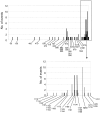
Amino acid position along CFH of all mutational events in non-Stx–HUS patients including published and present data. The x-axis indicates amino acid position.

References

    1. Ruggenenti P, Noris M, Remuzzi G. Thrombotic microangiopathy, hemolytic uremic syndrome, and thrombotic thrombocytopenic purpura. Kidney Int. 2001;60: 831-846. - PubMed
    1. Boyce TG, Swerdlow DL, Griffin PM. Escherichia coli O157:H7 and the hemolytic-uremic syndrome. N Engl J Med. 1995;333: 364-368. - PubMed
    1. Kaplan BS, Meyers KE, Schulman SL. The pathogenesis and treatment of hemolytic uremic syndrome. J Am Soc Nephrol. 1998;9: 1126-1133. - PubMed
    1. Schieppati A, Ruggenenti P, Cornejo RP, et al. Renal function at hospital admission as a prognostic factor in adult hemolytic uremic syndrome: The Italian Registry of Haemolytic Uremic Syndrome. J Am Soc Nephrol. 1992;2: 1640-1644. - PubMed
    1. Taylor CM, Chua C, Howie AJ, Risdon RA. Clinico-pathological findings in diarrhoea-negative haemolytic uraemic syndrome. Pediatr Nephrol. 2004;19: 419-425. - PubMed

Publication types

MeSH terms