. 2006 May;74(5):2568-77.
doi: 10.1128/IAI.74.5.2568-2577.2006.

Staphylococcus aureus escapes more efficiently from the phagosome of a cystic fibrosis bronchial epithelial cell line than from its normal counterpart

Affiliations

Staphylococcus aureus escapes more efficiently from the phagosome of a cystic fibrosis bronchial epithelial cell line than from its normal counterpart

Todd M Jarry et al. Infect Immun. 2006 May.

Abstract

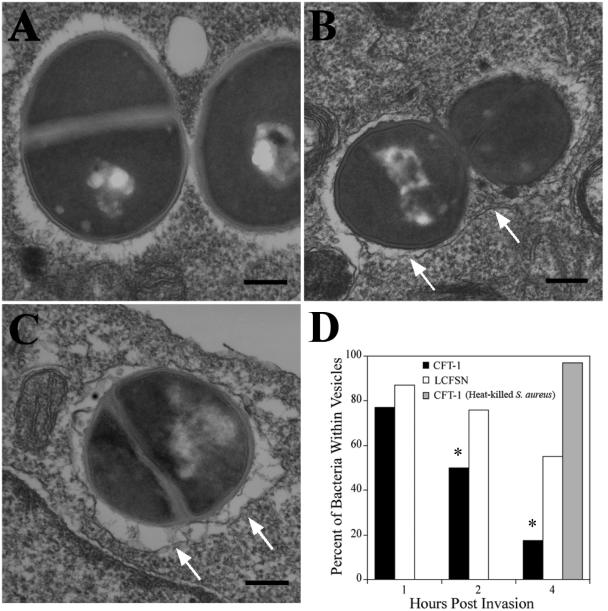
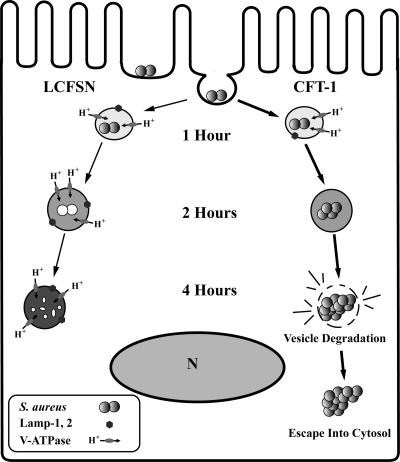
Staphylococcus aureus is frequently the initial bacterium isolated from young cystic fibrosis (CF) patients, and yet its role in CF disease progression has not been determined. Recent data from our lab demonstrates that S. aureus can invade and replicate within the CF tracheal epithelial cell line (CFT-1). Here we describe the finding that the fate of internalized S. aureus in CFT-1 cells differs from its complemented counterpart (LCFSN). S. aureus strain RN6390 was able to replicate within the mutant CFT-1 cells after invasion but not in the complemented LCFSN cells. At 1 h postinvasion, S. aureus containing vesicles within both cell lines acquired vacuolar-ATPase, lysosomal markers LAMP 1 and 2, and the lysomotrophic dye LysoTracker to a similar degree. However, at 4 h postinvasion, the percentage of S. aureus within CFT-1 cells associated with these markers decreased significantly compared to LCFSN, where the association approached 100%. Transmission electron microscopic analysis revealed that the majority of bacteria within CFT-1 cells were free in the cytosol at 4 h after invasion, whereas most S. aureus bacteria internalized by LCFSN cells remained within vesicles. These results demonstrate a fundamental difference in the fate of live S. aureus after invasion of CFT-1 versus LCFSN cell lines and may explain the propensity of S. aureus to cause chronic lung infection in CF patients.


Figures

FIG. 1.
FIG. 1.
Study of intracellular replication. (A) Intracellular replication of S. aureus in CFT-1 and LCFSN cells. Monolayers were infected with S. aureus strain RN6390 at an MOI of 10:1, followed by the addition of lysostaphin (10 μg/ml) 1 h postinfection to lyse extracellular S. aureus. The number of bacteria per cell in CFT-1 cells (circles) or complemented LCFSN cells (squares) was determined at hourly intervals by plating serial dilutions of cell lysates on tryptic soy agar plates. The bars indicate the mean ± the standard deviation (SD) of a representative experiment repeated three times. (B) Visual evidence of bacterial replication. Internalized S. aureus was visualized within CFT-1 or LCFSN 1 and 4 h postinvasion using the DNA stain To-Pro-3, which stains both epithelial and bacterial DNA. (C) Adherence and internalization of S. aureus to epithelial cells. Monolayers of CFT-1 or LCFSN cells were incubated with S. aureus on ice for 30 min. Infected monolayers were washed three times with cold PBS to remove unbound bacteria prior to CFU determination on Trypticase soy agar plates. We measured both adherent and internalized bacteria with these assays. Notably, internalized bacteria were also adherent prior to their uptake. (D) The number of internalized bacteria was determined by washing away nonadherent bacteria and incubated in the presence of lysostaphin for 30 min prior to CFU determination on Trypticase soy agar plates. (C and D) The data are shown on a log scale. The bars indicate the mean ± the SD of one representative experiment repeated three times.
FIG. 2.
FIG. 2.
Internalization of L. monocytogenes and B. subtilis into CFT-1 and LCFSN cells. Monolayers were infected with S. aureus, L. monocytogenes, or B. subtilis at an MOI of 10:1, followed by the addition of gentamicin (50 μg/ml) at 1 h postinfection to kill extracellular bacteria. Bacteria were enumerated on agar plates as described in Materials and Methods with CFT-1 (A) and LCFSN (B) cells. The bars indicate the mean ± the SD of one representative experiment repeated four times. Statistically significant differences determined using the Student t test (P < 0.05) were found between 2 and 6 h and are indicated by asterisks.
FIG. 3.
FIG. 3.
Analysis of S. aureus containing vesicles using the lysosomal markers LAMP-1 and LAMP-2. (A) Monolayers of epithelial cells grown on coverslips were infected with S. aureus at an MOI of 10:1 and processed for a 4-h time point as described in Materials and Methods. Infected cells were fixed and stained with anti-LAMP-1 (A and B) or anti-LAMP-2 (C and D) antibody visualized with Alexa Fluor 555-conjugated secondary antibody (green). Mammalian and bacterial DNA were stained with To-Pro-3 and visualized at 660 nm (red). S. aureus internalized by LCFSN cells were within LAMP-positive vesicles (arrows [A and C]), whereas a majority of bacteria internalized into CFT-1 cells lost their association with LAMP markers after 4 h (arrowheads). (E and F) Quantification of LAMP-1 and LAMP-2 association with bacteria, respectively. One hundred events were counted for each point on the graph. Each point represents the percentage of bacteria that was associated with LAMP-1 (E) or LAMP-2 (F) as determined by the presence of a positive ring around internalized bacteria at 1, 2, and 4 h postinfection. Each datum point represents the mean ± the standard error of the mean (SEM) of two experiments. The datum points differed significantly from the dead S. aureus control as marked by asterisks (P < 0.05). Bar, 10 μm.
FIG. 4.
FIG. 4.
z-series images of LCFSN and CFT-1 cells infected with S. aureus. LCFSN (A) and CFT-1 (B) cell lines were invaded with live S. aureus for 4 h, fixed, and then stained with LAMP-2 antibody (green) and To-Pro-3 to stain DNA (red). Each plane represents a 0.5-μm section starting at the basal side (0 μm) and moving toward the apical side (2.5 μm) of the cell. In both CFT-1 and LCFSN cell lines, bacteria could be found within LAMP-2-positive vesicles (arrows). However, in CFT-1 cells, most bacteria did not reside within LAMP-2-positive vesicles (arrowheads).
FIG. 5.
FIG. 5.
Analysis of lysosomal acidification. Monolayers of epithelial cells grown on coverslips were infected with S. aureus at an MOI of 10:1. Thirty minutes prior to each time point, LysoTracker (LyT) was added to the cell culture media to label acidic vesicles prior to fixing at 1, 2, and 4 h postinfection. (A and B) A majority of S. aureus within CFT-1 cells lost their association with LyT after 4 h (arrowheads) (B), whereas S. aureus within LCFSN cells maintained their association with LyT (arrows) (A). (C) Quantitation of bacteria that colocalized with LyT. One hundred events are recorded for each time point. Each point represents the percentage of internalized bacteria associated with the acidic marker LyT. Each datum point represents the mean ± the SEM of two experiments. The datum points significantly different from the dead S. aureus control are marked by asterisks (P < 0.001). Bar, 10 μm.
FIG. 6.
FIG. 6.
Association of S. aureus-containing vesicles with V-ATPase. Monolayers of LCFSN (A) and CFT-1 (B) epithelial cells were infected with live S. aureus at an MOI of 10:1. Infected cells were fixed and stained with anti-V-ATPase antibody visualized with Alexa Fluor 555-conjugated secondary antibody (green). Mammalian and bacterial DNA were stained with To-Pro-3 and visualized at 660 nm (red). S. aureus bacteria within LCFSN cells were stained V-ATPase positive after 4 h of internalization (C, ▪), while the number of S. aureus within CFT-1 cells associated with V-ATPase decreased over the time (C, ○). The events were quantitated as described for Fig. 5C. Each datum point represents the mean ± the SEM of two experiments. Bar, 10 μm.
FIG. 7.
FIG. 7.
EM analysis of S. aureus-infected CFT-1 and LCFSN cells. Monolayers of CFT-1 or LCFSN cells were grown in six-well plates and infected with either heat-killed or live S. aureus. Samples were then fixed and stained for subsequent transmission EM analysis as described in Materials and Methods. (A to C) At 4 h postinfection, heat-killed bacteria within CFT-1 cells (B) and most live S. aureus within LCFSN cells (C) were found in membrane-bound vesicles (arrows), while live S. aureus within CFT-1 cells (A) were found free in the host cell cytoplasm. Bar, 200 nm. (D) The internalized bacteria that were either within a vesicle or free in the cytoplasm were quantified by counting 100 events on a series of EM images. Heat-killed bacteria were also included as an additional control at the 4-h time point. The datum points significantly different from the 1-h time point are marked by asterisks (P < 0.05). Bar, 10 μm.
FIG. 8.
FIG. 8.
Proposed model of S. aureus trafficking within CFT-1 cell line.

References

    1. Bals, R., D. J. Weiner, and J. M. Wilson. 1999. The innate immune system in cystic fibrosis lung disease. J. Clin. Investig. 103:303-307. - PMC - PubMed
    1. Bayles, K. W., C. A. Wesson, L. E. Liou, L. K. Fox, G. A. Bohach, and W. R. Trumble. 1998. Intracellular Staphylococcus aureus escapes the endosome and induces apoptosis in epithelial cells. Infect. Immun. 66:336-342. - PMC - PubMed
    1. Beauregard, K. E., K. D. Lee, R. J. Collier, and J. A. Swanson. 1997. pH-dependent perforation of macrophage phagosomes by listeriolysin O from Listeria monocytogenes. J. Exp. Med. 186:1159-1163. - PMC - PubMed
    1. Burns, J. L., J. Emerson, J. R. Stapp, D. L. Yim, J. Krzewinski, L. Louden, B. W. Ramsey, and C. R. Clausen. 1998. Microbiology of sputum from patients at cystic fibrosis centers in the United States. Clin. Infect. Dis. 27:158-163. - PubMed
    1. Burton, P. R., N. Kordova, and D. Paretsky. 1971. Electron microscopic studies of the rickettsia Coxiella burnetii: entry, lysosomal response, and fate of rickettsial DNA in L-cells. Can. J. Microbiol. 17:143-150. - PubMed

Publication types

MeSH terms

Substances