. 2006 May;74(5):2578-86.
doi: 10.1128/IAI.74.5.2578-2586.2006.

The NKG2D-activating receptor mediates pulmonary clearance of Pseudomonas aeruginosa

Affiliations

The NKG2D-activating receptor mediates pulmonary clearance of Pseudomonas aeruginosa

Michael T Borchers et al. Infect Immun. 2006 May.

Abstract

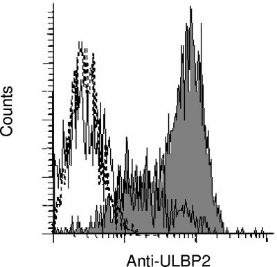
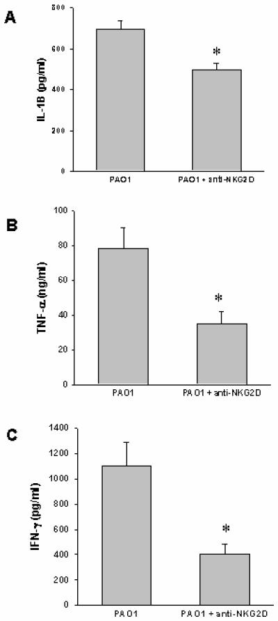
The NKG2D-activating receptor is expressed on cytotoxic lymphocytes and interacts with ligands expressed on the surface of cells stressed by pathogenic and nonpathogenic stimuli. In this study, we investigated the physiologic importance of NKG2D receptor-ligand interactions in response to acute pulmonary Pseudomonas aeruginosa infection. P. aeruginosa infection increased the expression of mouse NKG2D ligands (Rae1) in airway epithelial cells and alveolar macrophages in vivo and also increased the cell surface expression of human NKG2D ligands (ULBP2) on airway epithelial cells in vitro. NKG2D receptor blockade with a specific monoclonal antibody inhibited the pulmonary clearance of P. aeruginosa. NKG2D receptor blockade also resulted in decreased production of Th1 cytokines and nitric oxide in the lungs of P. aeruginosa-infected mice. Additionally, NKG2D receptor blockade reduced the epithelial cell sloughing that accompanies P. aeruginosa infection. Macrophage phagocytosis and bronchoalveolar lavage cellularity were not different in P. aeruginosa-infected mice with and without NKG2D receptor blockade. These results demonstrate the importance of NKG2D-mediated immune activation in the clearance of acute bacterial infection and suggest that epithelial cell-lymphocyte interactions mediate pulmonary cytokine production, epithelial cell integrity, and bacterial clearance.


Figures

FIG. 1.
FIG. 1.
NKG2D ligands are induced in the lungs of mice infected with P. aeruginosa. (A) Western blot analysis of RAE-1 in whole lungs of mice exposed to ∼1 × 10⁶ CFU of P. aeruginosa for 0, 8, and 24 h. The blot is representative of results obtained with four to six mice per group. (B to E) NKG2D ligand expression in lung epithelial cells and alveolar macrophages of mice exposed to ∼1 × 10⁶ CFU of P. aeruginosa for 24 h. The accumulation of NKG2D ligands in the lungs of CF1 mice was assessed by immunohistochemistry analysis using paraffin-embedded sections and a goat polyclonal antibody against RAE-1γ. (B) Large airway of PBS-treated mouse. (C) Large airway of P. aeruginosa-infected mouse. (D) Lung parenchyma of PBS-treated mouse. (E) Lung parenchyma of P. aeruginosa-infected mouse. The photomicrographs are representative of five mice per group. Original magnification, ×400.
FIG. 2.
FIG. 2.
Expression and induction of an NKG2D ligand on the surface of human airway epithelial cells. A simian virus 40-transformed primary tracheal epithelial cell line (9HTEo-) was treated with P. aeruginosa (multiplicity of infection, 10) for 24 h. The dashed line histograms indicate the results for isotype controls, the solid line histograms indicate the results for PBS-treated cells, and the shaded histograms indicate the results for P. aeruginosa-infected cells. ULBP2 cell surface expression was assessed by immunofluorescence and fluorescence-activated cell sorting analysis using mouse monoclonal antibodies, followed by phycoerythrin-conjugated goat anti-mouse antibodies. The histograms are representative of three independent experiments.
FIG. 3.
FIG. 3.
NKG2D receptor expression on pulmonary lymphocytes. NKG2D-expressing lymphocyte populations in perfused, digested lungs were analyzed by flow cytometry. Cells were purified as described in Materials and Methods and were labeled with the following antibodies: phycoerythrin-conjugated isotype control (A), phycoerythrin-conjugated NKG2D (B), allophycocyanin-conjugated NK1.1 and phycoerythrin-conjugated NKG2D (C), and phycoerythrin-conjugated NKG2D with allophycocyanin-conjugated CD8 antibodies (D). Dot plot diagrams were derived from the lymphocyte gate ascertained from forward and side scatter plots. The dot plots are representative of three independent experiments. APC, allophycocyanin; PE, phycoerythrin.
FIG. 4.
FIG. 4.
NKG2D signaling blockade inhibits the pulmonary clearance of P. aeruginosa. Mice were treated with the NKG2D receptor blocking antibody (100 μg of CX5) or control IgG prior to P. aeruginosa infection, and the bacterial CFU in the lungs were assessed at 24 h. Larger amounts of P. aeruginosa were recovered from the lungs of mice that were treated with the CX5 antibody. The data are means and standard errors of the means for seven or eight mice per group. The asterisk indicates that the value was significantly greater than the value for P. aeruginosa-infected, control, IgG-treated mice (P < 0.05).
FIG. 5.
FIG. 5.
NKG2D signaling blockade does not impair AM phagocytosis of P. aeruginosa. Mice were treated with the NKG2D receptor blocking antibody (100 μg of CX5) or control IgG prior to GFP-PAO1 infection, and the percentages of AM in the bronchoalveolar lavage that had ingested bacteria were determined by fluorescence microscopy. The data are means and standard errors of the means for three mice per group.
FIG. 6.
FIG. 6.
NKG2D signaling mediates cytokine production in response to P. aeruginosa infection. Mice were treated with the NKG2D receptor blocking antibody (100 μg of CX5) or control IgG prior to P. aeruginosa infection, and cytokine levels in the whole lung were determined by an enzyme-linked immunosorbent assay at 24 h. Smaller amounts of IL-1β (A), TNF-α (B), and IFN-γ (C) were detected in the lungs of mice that were treated with the CX5 antibody. The data are means and standard errors of the means for five or six mice per group. An asterisk indicates that the value was significantly less than the value for P. aeruginosa-infected, control, IgG-treated mice (P < 0.05).
FIG. 7.
FIG. 7.
NKG2D signaling mediates nitric oxide (NO) production in response to P. aeruginosa infection. Mice were treated with the NKG2D receptor blocking antibody (100 μg of CX5) or control IgG prior to P. aeruginosa infection, and nitrite levels in the BAL were determined by the Griess reaction at 8 and 24 h postinfection. The data are means and standard errors of the means for five or six mice per group. The asterisk indicates that the value was significantly less than the value for P. aeruginosa-infected, control, IgG-treated mice (P < 0.05).
FIG. 8.
FIG. 8.
Role of NKG2D in pulmonary inflammation and epithelial cell injury in response to P. aeruginosa infection. (A) Mice were treated with the NKG2D receptor blocking antibody (100 μg of CX5) or control IgG prior to P. aeruginosa infection, and BAL cellularity was assessed at 24 h. No significant differences were observed in the total leukocytes recovered or the composition of the leukocytes in mice treated with the CX5 antibody. (B) The percentage of epithelial cells recovered from the BAL fluid at 24 h was reduced in P. aeruginosa-infected mice treated with the CX5 antibody compared to control-treated mice. The data are means and standard errors of the means for five mice per group. The asterisk indicates that the value was significantly less than the value for P. aeruginosa-infected, control, IgG-treated mice (P < 0.05).

References

    1. Agerberth, B., J. Charo, J. Werr, B. Olsson, F. Idali, L. Lindbom, R. Kiessling, H. Jornvall, H. Wigzell, and G. H. Gudmundsson. 2000. The human antimicrobial and chemotactic peptides LL-37 and alpha-defensins are expressed by specific lymphocyte and monocyte populations. Blood 96:3086-3093. - PubMed
    1. Bacon, L., R. A. Eagle, M. Meyer, N. Easom, N. T. Young, and J. Trowsdale. 2004. Two human ULBP/RAET1 molecules with transmembrane regions are ligands for NKG2D. J. Immunol. 173:1078-1084. - PubMed
    1. Bauer, S., V. Groh, J. Wu, A. Steinle, J. H. Phillips, L. L. Lanier, and T. Spies. 1999. Activation of NK cells and T cells by NKG2D, a receptor for stress-inducible MICA. Science 285:727-729. - PubMed
    1. Buret, A., M. L. Dunkley, G. Pang, R. L. Clancy, and A. W. Cripps. 1994. Pulmonary immunity to Pseudomonas aeruginosa in intestinally immunized rats: roles of alveolar macrophages, tumor necrosis factor alpha, and interleukin-1 alpha. Infect. Immun. 62:5335-5343. - PMC - PubMed
    1. Calum, H., C. Moser, P. O. Jensen, R. Shirai, and N. Hoiby. 2003. Cytokine and surface receptor diversity of NK cells in resistant C3H/HeN and susceptible BALB/c mice with chronic Pseudomonas aeruginosa lung infection. APMIS 111:891-897. - PubMed

Publication types

LinkOut - more resources