. 2006 May;74(5):2628-36.
doi: 10.1128/IAI.74.5.2628-2636.2006.

The most polymorphic residue on Plasmodium falciparum apical membrane antigen 1 determines binding of an invasion-inhibitory antibody

Affiliations

The most polymorphic residue on Plasmodium falciparum apical membrane antigen 1 determines binding of an invasion-inhibitory antibody

A M Coley et al. Infect Immun. 2006 May.

Abstract

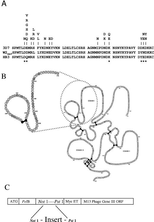
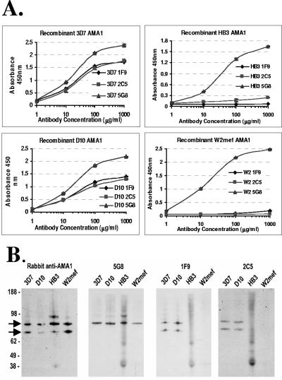
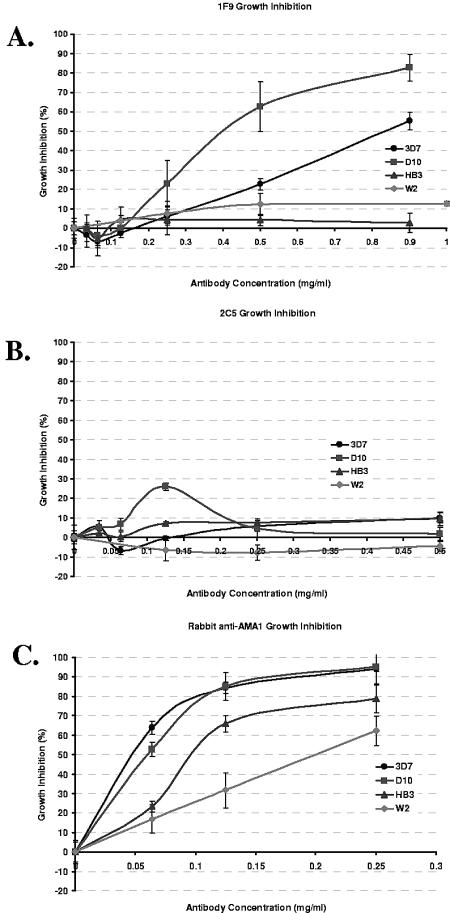
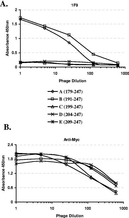
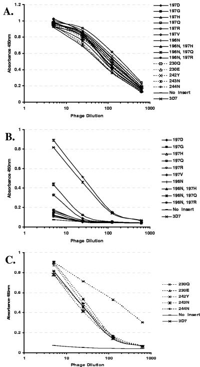
Apical membrane antigen 1 (AMA1) is currently one of the leading malarial vaccine candidates. Anti-AMA1 antibodies can inhibit the invasion of erythrocytes by Plasmodium merozoites and prevent the multiplication of blood-stage parasites. Here we describe an anti-AMA1 monoclonal antibody (MAb 1F9) that inhibits the invasion of Plasmodium falciparum parasites in vitro. We show that both reactivity of MAb 1F9 with AMA1 and MAb 1F9-mediated invasion inhibition were strain specific. Site-directed mutagenesis of a fragment of AMA1 displayed on M13 bacteriophage identified a single polymorphic residue in domain I of AMA1 that is critical for MAb 1F9 binding. The identities of all other polymorphic residues investigated in this domain had little effect on the binding of the antibody. Examination of the P. falciparum AMA1 crystal structure localized this residue to a surface-exposed alpha-helix at the apex of the polypeptide. This description of a polymorphic inhibitory epitope on AMA1 adds supporting evidence to the hypothesis that immune pressure is responsible for the polymorphisms seen in this molecule.


Figures

FIG. 1.
FIG. 1.
(A) Alignment of the 3D7 and D10 AMA1 MAb 1F9 minimally reactive fragment amino acid sequence with the corresponding sequences from HB3 and W2mef. The polymorphisms present in the database are indicated above the sequences with the amino acid alternatives identified at those positions. Asterisks indicate the positions of the individual amino acid substitutions carried out in this study. (B) Primary “string-of-beads” representation of 3D7 AMA1, modified from that of Hodder et al. (19), with the relative position of the MAb 1F9-reactive fragment indicated. (C) Diagrammatic representation of the cloning approach taken in order to display AMA1 domain I and fragments thereof on M13 bacteriophage. The AMA1 sequences were ligated in frame into pHENH6 phagemid vector. Kunkel mutagenesis was carried out on single-stranded DNA derived from these ligation products.
FIG. 2.
FIG. 2.
(A) ELISA analysis of MAb 1F9, 5G8, 2C5 reactivity with recombinant refolded AMA1 derived from P. falciparum strains 3D7, D10, HB3, and W2mef. AMA1 preparations were adsorbed onto the wells, and antibodies were then allowed to react at the indicated concentrations. Absorbances at 450 nm and MAb concentrations are indicated on the ordinate and abscissa, respectively. (B) Western analysis of AMA1 expressed by parasite strains 3D7, D10, HB3, and W2mef. Schizont-stage parasites were separated by SDS-PAGE and blotted onto polyvinylidene difluoride, and blots were visualized with MAbs 1F9, 2C5, and 5G8 and rabbit anti-AMA1. Arrows indicate the locations of the unprocessed 83-kDa and processed 66-kDa bands.
FIG. 3.
FIG. 3.
Growth inhibition assays of 3D7, D10, HB3, and W2mef parasites. Each antibody, i.e., 1F9 (A), 2C5 (B), and rabbit anti-AMA1 (C), was assayed for its growth inhibition qualities. Data points indicate the means of three identical wells, and error bars indicate ± one standard deviation from the mean.
FIG. 4.
FIG. 4.
Defining the MAb 1F9 minimally reactive fragment of 3D7 AMA1. Fragment A represents amino acids 179 to 247, fragment B represents amino acids 191 to 247, fragment C represents amino acids 199 to 247, fragment D represents amino acids 204 to 247, and fragment E represents amino acids 209 to 247 of 3D7 AMA1 displayed on phage. Each fragment was assayed for its reactivity with the anti-Myc epitope tag MAb 9E10 (A) and MAb 1F9 (B).
FIG. 5.
FIG. 5.
ELISA analysis of point mutants of 3D7 AMA1 domain I displayed on phage. MAbs 9E10 (A) and 1F9 (B and C) were adsorbed to the plate and the phage added at indicated dilutions. Reactivity of the phage with MAb 9E10 indicates the relative level of AMA1 domain I in each preparation.
FIG.6.
FIG.6.
X-ray structure of P. falciparum 3D7 AMA1 domains I and II. (A) The intact domains I and II, with the 57-residue MAb 1F9-reactive fragment represented by the solid surface. Residue 197 is represented in blue. The mottled gray surface represents the remainder of AMA1 domains I and II, which is not necessary for reactivity with MAb 1F9. Other polymorphic residues studied (residues 196, 230, 242, 243, and 244) are represented in yellow. The disulfide-bonded cysteines 217 and 247 are represented in orange. (B) Ribbon representation of the MAb 1F9-reactive fragment in the same orientation and with the same residue color assignments as in panel A. (C) Reverse-angle orientation demonstrating the relative locations of the critical 1F9 residue 197 and the surface-exposed residues critical for 4G2dc1 binding. Residue 197 (blue) is diametrically opposite the 4G2dc1 critical residues (red). The 57-residue MAb 1F9-reactive fragment is represented by the pale blue surface, and the hydrophobic cleft is represented in yellow.

Similar articles

Cited by

References

    1. Anders, R. F., P. E. Crewther, S. Edwards, M. Margetts, M. L. Matthew, B. Pollock, and D. Pye. 1998. Immunisation with recombinant AMA-1 protects mice against infection with Plasmodium chabaudi. Vaccine 16:240-247. - PubMed
    1. Anders, R. F., D. J. McColl, and R. L. Coppel. 1993. Molecular variation in Plasmodium falciparum: polymorphic antigens of asexual erythrocytic stages. Acta Trop. 53:239-253. - PubMed
    1. Bai, T., M. Becker, A. Gupta, P. Strike, V. J. Murphy, R. F. Anders, and A. H. Batchelor. 2005. Structure of AMA1 from Plasmodium falciparum reveals a clustering of polymorphisms that surround a conserved hydrophobic pocket. Proc. Natl. Acad. Sci. USA 102:12736-12741. - PMC - PubMed
    1. Basco, L. K., F. Marquet, M. M. Makler, and J. Le Bras. 1995. Plasmodium falciparum and Plasmodium vivax: lactate dehydrogenase activity and its application for in vitro drug susceptibility assay. Exp. Parasitol. 80:260-271. - PubMed
    1. Casey, J. L., A. M. Coley, R. F. Anders, V. J. Murphy, K. S. Humberstone, A. W. Thomas, and M. Foley. 2004. Antibodies to malaria peptide mimics inhibit Plasmodium falciparum invasion of erythrocytes. Infect. Immun. 72:1126-1134. - PMC - PubMed

Publication types

LinkOut - more resources