. 2006 May;74(5):2760-6.
doi: 10.1128/IAI.74.5.2760-2766.2006.

In vivo activation of naive CD4+ T cells in nasal mucosa-associated lymphoid tissue following intranasal immunization with recombinant Streptococcus gordonii

Affiliations

In vivo activation of naive CD4+ T cells in nasal mucosa-associated lymphoid tissue following intranasal immunization with recombinant Streptococcus gordonii

Donata Medaglini et al. Infect Immun. 2006 May.

Abstract

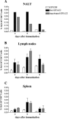
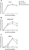
The antigen-specific primary activation of CD4+ T cells was studied in vivo by adoptive transfer of ovalbumin-specific transgenic T cells (KJ1-26+ CD4+) following intranasal immunization with recombinant Streptococcus gordonii. A strain of S. gordonii expressing on its surface a model vaccine antigen fused to the ovalbumin (OVA) peptide from position 323 to 339 was constructed and used to study the OVA-specific T-cell activation in nasal mucosa-associated lymphoid tissue (NALT), lymph nodes, and spleens of mice immunized by the intranasal route. The recombinant strain, but not the wild type, activated the OVA-specific CD4+ T-cell population in the NALT (89% of KJ1-26+ CD4+ T cells) just 3 days following immunization. In the cervical lymph nodes and in the spleen, the percentage of proliferating cells was initially low, but it reached the peak of activation at day 5 (90%). This antigen-specific clonal expansion of KJ1-26+ CD4+ T cells after intranasal immunization was obtained with live and inactivated recombinant bacteria, and it indicates that the NALT is the site of antigen-specific T-cell priming.


Figures

FIG. 1.
FIG. 1.
Fusion protein expressed on the surface of recombinant S. gordonii GP1422. A. Schematic structure of the fusion protein displayed on the surface of S. gordonii GP1422. The recombinant protein is constituted by the first N-terminal 122 aa and the last C-terminal 140 aa of M6, OVA323-339 (ISQAVHAAHAEINEAGR), P27 protein (375 aa), and the E-tag (GAPVPYPDPLEPR). B. Western blot analysis of cell surface proteins of recombinant S. gordonii GP1422. Samples were reacted with anti-M6 polyclonal antibodies. Lane 1, control strain GP1295; lane 2, recombinant GP1422. C. Flow cytometric analysis of S. gordonii GP1422 (solid histogram) and control strain GP1295 (open histogram). Bacterial cells were reacted with anti-P27 rabbit serum and then with FITC-conjugated anti-rabbit IgG.
FIG. 2.
FIG. 2.
KJ1-26+ CD4+ T-cell expansion in secondary lymphoid organs following intranasal immunization with live and inactivated recombinant S. gordonii. BALB/c mice were adoptively transferred with DO11.10 T cells and then immunized by the intranasal route with 109 CFU of either the control strain GP1295 or live or inactivated OVA-expressing GP1422. NALT, cervical lymph nodes, and spleens were collected from mice at 3, 5, and 8 days following immunization. At each time point, three mice were sacrificed for each group and analyzed either as single samples (lymph nodes and spleen) or as a pool (NALT). DO11.10 T cells were identified using PE-conjugated anti-clonotypic KJ1-26 and CyChrome-conjugated anti-CD4 monoclonal antibodies. The percentage of KJ1-26+ CD4+ T cells for each organ is reported (mean ± standard deviation).
FIG. 3.
FIG. 3.
Clonal expansion of KJ1-26+ CD4+ T cells in lymphoid organs 5 days following intranasal immunization with S. gordonii. BALB/c mice were adoptively transferred with CFSE-labeled DO11.10 T cells and then immunized by the intranasal route with 109 CFU of either control strain GP1295 or live or inactivated OVA-expressing strain GP1422. NALT, cervical lymph nodes, and spleens were collected at day 5 from three mice per group and analyzed as single samples (lymph nodes and spleen) or as a pool (NALT). CFSE dilution was analyzed on the gated KJ1-26+ CD4+ population with light scatter properties of lymphocytes. The M1 marker above the peaks was used to calculate the percentage of proliferating cells. n.d, not detectable.
FIG. 4.
FIG. 4.
Time course analysis of the clonal expansion of KJ1-26+ CD4+ T cells following intranasal immunization with recombinant S. gordonii. The clonal expansion of KJ1-26+ CD4+ T cells following intranasal immunization with 109 CFU of live recombinant GP1422, inactivated recombinant GP1422, or control strain GP1295 was analyzed at different time points (3, 5, and 8 days postimmunization) in NALT, cervical lymph nodes, and spleens of BALB/c mice adoptively transferred with CFSE-labeled transgenic T cells. DO11.10 T cells were identified using PE-conjugated anti-clonotypic KJ1-26 and CyChrome-conjugated anti-CD4 monoclonal antibodies. The percentage of proliferating KJ1-26+ CD4+ T cells is reported as the mean ± standard deviation for three animals for lymph nodes and spleens and as a pool for NALT cells. KJ1-26+ CD4+ T cells were undetectable in the NALT of mice immunized with control strain GP1295.

References

    1. Asanuma, H., A. H. Thompson, T. Iwasaki, Y. Sato, Y. Inaba, C. Aizawa, T. Kurata, and S. Tamura. 1997. Isolation and characterization of mouse nasal-associated lymphoid tissue. J. Immunol. Methods 202:123-131. - PubMed
    1. Bergquist, C., E. L. Johansson, T. Lagergard, J. Holmgreen, and A. Rudin. 1997. Intranasal vaccination of humans with recombinant cholera toxin B subunit induces systemic and local antibody responses in the upper respiratory tract and the vagina. Infect. Immun. 65:2676-2684. - PMC - PubMed
    1. Bienenstock, J., and M. R. McDermott. 2005. Bronchus- and nasal-associated lymphoid tissue. Immunol. Rev. 206:22-31. - PubMed
    1. Corinti, S., D. Medaglini, A. Cavani, M. Rescigno, G. Pozzi, P. Ricciardi-Castagnoli, and G. Girolomoni. 1999. Human dendritic cells very efficiently present a heterologous antigen expressed on the surface of recombinant Gram-positive bacteria to CD4+ T lymphocytes. J. Immunol. 163:3029-3036. - PubMed
    1. Corinti, S., D. Medaglini, C. Prezzi, G. Pozzi, and G. Girolomoni. 2000. Human dendritic cells are superior to B cells at presenting a major histocompatibility complex class-II restricted heterologous antigen expressed on recombinant Streptococcus gordonii. Infect. Immun. 68:1879-1883. - PMC - PubMed

Publication types