. 2006 May;74(5):2839-48.
doi: 10.1128/IAI.74.5.2839-2848.2006.

Murine malaria infection induces fetal loss associated with accumulation of Plasmodium chabaudi AS-infected erythrocytes in the placenta

Affiliations

Murine malaria infection induces fetal loss associated with accumulation of Plasmodium chabaudi AS-infected erythrocytes in the placenta

Jayakumar Poovassery et al. Infect Immun. 2006 May.

Abstract

Malarial infection in nonimmune women is a risk factor for pregnancy loss, but the role that maternal antimalarial immune responses play in fetal compromise is not clear. We conducted longitudinal and serial sacrifice studies to examine the pathogenesis of malaria during pregnancy using the Plasmodium chabaudi AS/C57BL/6 mouse model. Peak parasitemia following inoculation with 1,000 parasite-infected murine erythrocytes and survival were similar in infected pregnant and nonpregnant mice, although development of parasitemia and anemia was slightly accelerated in pregnant mice. Importantly, pregnant mice failed to maintain viable pregnancies, most aborting before day 12 of gestation. At abortion, maternal placental blood parasitemia was statistically significantly higher than peripheral parasitemia. Infected mice had similar increases in spleen size and cellularity which were statistically significantly higher than in uninfected mice. In contrast, splenocyte proliferation in response to mitogenic stimulation around peak parasitemia was statistically significantly reduced in both groups of infected mice compared to uninfected, nonpregnant mice, suggesting that lymphoproliferation is not a good indicator of the antimalarial immune responses in pregnant or nonpregnant animals. This study suggests that while pregnant and nonpregnant C57BL/6 mice are equally capable of mounting an effective immune response to and surviving P. chabaudi AS infection, pregnant mice cannot produce viable pups. Fetal loss appears to be associated with placental accumulation of infected erythrocytes. Further study is required to determine to what extent maternal antimalarial immune responses, anemia, and placental accumulation of parasites contribute to compromised pregnancy in this model.


Figures
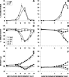
FIG. 1.
FIG. 1.
Parasitemia, hematocrits, and weight changes in longitudinal and serial sacrifice studies of P. chabaudi AS-infected and control B6 mice. Eight- to 9-week-old C57BL/6 mice were injected intravenously with 1,000 P. chabaudi AS-infected erythrocytes or 200 μl of phosphate-buffered saline (control mice) per 20 g of body weight on GD/ED 0. Percent parasitemia (A and B) from Giemsa-stained thin smears, hematocrits (C and D) of tail vein blood, and weights (E and F) were assessed at 1- to 2-day intervals as shown. Mice were divided into four groups: UNP, INP, UP, IP. Groups were either followed longitudinally for 18 days (n = 3 for all groups; A, C, and E) or sacrificed at days 6, 8, 9, 10, 11, or 12 in two serial sacrifice studies (B, D, and F). Although IP mice aborted, resorbed fetuses, or had only dead embryos in their uteri by GD 12, for the sake of clarity and illustration of the point, mice in this group were retained as such for the whole of the longitudinal experiment. For serial sacrifice studies, clinical parameters were measured in mice as described in Materials and Methods until the day of sacrifice. Starting sample sizes were as follows: UNP, 9; INP, 39; UP, 56; IP, 67. Number of mice sacrificed (INP, UP, and IP, respectively) at each GD/ED was as follows: at GD/ED 6, n = 8, 12, and 14 sacrificed; at GD/ED 8, n = 5, 10, and 8; at GD/ED 9, n = 8, 13, and 13; at GD/ED 10, n = 5, 5, and 14; at GD/ED 11, n = 5, 9, and 11; at GD/ED 12, n = 8, 7, 7. Three UNP mice were sacrificed at ED 6, 9, and 12. All data presented are means ± standard errors of the means. The y axes in panels E and F begin at 15 g to avoid compression and poor visualization of the data. Statistical differences (all P < 0.05; Proc GLM, Tukey) in the longitudinal study were found for the following comparisons: hematocrit at GD/ED 6 (UP versus UNP), GD/ED 10 (IP versus uninfected), GD/ED 12 and14 (infected versus uninfected), GD/ED 16 (IP versus UNP), and GD/ED 18 (INP versus IP and UNP); weight at GD/ED 6 (UNP versus UP), GD/ED 7 and 8 (UNP and INP versus UP), and GD/ED 10 to 18 (all groups versus UP). In the serial sacrifice studies, statistical analysis results included the following significant differences (P < 0.05 unless otherwise noted): parasitemia at GD/ED 8 (INP versus IP; P = 0.0004 based on Welch's analysis of variance); hematocrit at GD/ED 8 to 10 (IP versus all groups), GD/ED 10 (INP versus all groups), GD/ED 11 (infected versus UP), and GD/ED 12 (infected versus uninfected); weight at GD/ED 6 and 8 (INP versus IP and UP), GD/ED 7 (INP versus all groups), GD/ED 9 (IP versus INP), GD/ED 9 to 12 (infected versus UP), and GD/ED 11 and 12 (INP versus UNP).
FIG. 2.
FIG. 2.
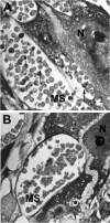
P. chabaudi AS-infected erythrocytes in the placenta of an aborting mouse. (A) Giemsa-stained placental section (2 μm thick) from an IP mouse undergoing abortion on GD/ED 11, showing infected erythrocytes in the maternal blood spaces (arrows). (B) Giemsa-stained placental section from a UP mouse at the same time point, showing normal RBCs in the maternal sinusoids. MS, maternal sinusoid; N, giant cell nucleus. Photographs were prepared using Adobe Photoshop version 8.0.
FIG. 3.
FIG. 3.
Comparison of placental and peripheral parasitemias in aborting and nonaborting IP mice. Placental parasitemias were scored in Giemsa-stained, 2-μm-thick placental sections by counting at least 1,000 RBCs in the maternal blood spaces. Peripheral parasitemias were determined in Giemsa-stained tail blood smears. n = 5 at each time point per group except for day 9 (n = 3). *, P = 0.003, Student's t test.
FIG. 4.
FIG. 4.
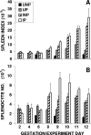
Changes in spleen index and cell number in experimental mice. Mice were experimentally manipulated as indicated in the legends for Fig. 1 and 2. (A) Spleen index, calculated as a proportion of spleen weight to total body weight. (B) Cell number, calculated from isolated splenocytes enumerated by trypan blue exclusion. Data were pooled from two replicate experiments and are presented as means ± standard errors of the means of 5 to 10 mice for the IP, INP, and UP groups (n = 4, IP day 12) and 3 UNP mice analyzed individually per time point. Statistically significant differences (P < 0.05, Proc GLM and Tukey) in the spleen indexes were noted for the following comparisons: day 6 (UNP versus IP and UP), day 8 (UP versus IP), days 9 and 12 (both UNP and UP versus all groups), and days 10 and 11 (UP versus IP and INP). For spleen cell number, significant differences (P < 0.05) were found for the following comparisons (based on Proc GLM and Tukey unless otherwise noted): day 6 (for INP and UNP versus UP and for UNP versus IP), day 8 (UP versus INP; Dunnett's T3), day 9 (for UNP versus INP and for IP versus all groups); day 11 (IP versus UP; Dunnett's T3), day 12 (UNP versus all groups).
FIG. 5.
FIG. 5.
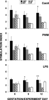
Proliferative response of spleen cells in experimental mice. Spleen cells from P. chabaudi AS-infected pregnant, infected nonpregnant, and uninfected controls collected aseptically on the days indicated were cultured with ConA (2 μg/ml), PWM (2 μg/ml), and LPS (1 μg/ml) or medium as the control. BrdU uptake was measured, and data shown are means ± standard errors of the means for three mice per group. *, P < 0.05 for INP and IP versus UNP; **, P <0.05 for INP versus UNP (Proc GLM and Duncan).

Similar articles

Cited by

References

    1. Abdalla, S., and D. J. Weatherall. 1982. The direct antiglobulin test in P. falciparum malaria. Br. J. Haematol. 51:415-425. - PubMed
    1. Abdalla, S., D. J. Weatherall, S. N. Wickramasinghe, and M. Hughes. 1980. The anaemia of P. falciparum malaria. Br. J. Haematol. 46:171-183. - PubMed
    1. Anonymous. 1997. World malaria situation in 1994. Wkly. Epidemiol. Rec. 72:269-276. - PubMed
    1. Beeson, J. G., G. V. Brown, M. E. Molyneux, C. Mhango, F. Dzinjalamala, and S. J. Rogerson. 1999. Plasmodium falciparum isolates from infected pregnant women and children are associated with distinct adhesive and antigenic properties. J. Infect. Dis. 180:464-472. - PMC - PubMed
    1. Beier, J. C., C. N. Oster, F. K. Onyango, J. D. Bales, J. A. Sherwood, P. V. Perkins, D. K. Chumo, D. V. Koech, R. E. Whitmire, C. R. Roberts, et al. 1994. Plasmodium falciparum incidence relative to entomologic inoculation rates at a site proposed for testing malaria vaccines in western Kenya. Am. J. Trop. Med. Hyg. 50:529-536. - PubMed

Publication types