Attenuated bioluminescent Brucella melitensis mutants GR019 (virB4), GR024 (galE), and GR026 (BMEI1090-BMEI1091) confer protection in mice
- PMID: 16622231
- PMCID: PMC1459743
- DOI: 10.1128/IAI.74.5.2925-2936.2006
Attenuated bioluminescent Brucella melitensis mutants GR019 (virB4), GR024 (galE), and GR026 (BMEI1090-BMEI1091) confer protection in mice
Abstract
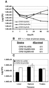
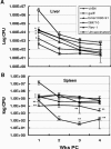
In vivo bioluminescence imaging is a persuasive approach to investigate a number of issues in microbial pathogenesis. Previously, we have applied bioluminescence imaging to gain greater insight into Brucella melitensis pathogenesis. Endowing Brucella with bioluminescence allowed direct visualization of bacterial dissemination, pattern of tissue localization, and the contribution of Brucella genes to virulence. In this report, we describe the pathogenicity of three attenuated bioluminescent B. melitensis mutants, GR019 (virB4), GR024 (galE), and GR026 (BMEI1090-BMEI1091), and the dynamics of bioluminescent virulent bacterial infection following vaccination with these mutants. The virB4, galE, and BMEI1090-BMEI1091 mutants were attenuated in interferon regulatory factor 1-deficient (IRF-1(-/-)) mice; however, only the GR019 (virB4) mutant was attenuated in cultured macrophages. Therefore, in vivo imaging provides a comprehensive approach to identify virulence genes that are relevant to in vivo pathogenesis. Our results provide greater insights into the role of galE in virulence and also suggest that BMEI1090 and downstream genes constitute a novel set of genes involved in Brucella virulence. Survival of the vaccine strain in the host for a critical period is important for effective Brucella vaccines. The galE mutant induced no changes in liver and spleen but localized chronically in the tail and protected IRF-1(-/-) and wild-type mice from virulent challenge, implying that this mutant may serve as a potential vaccine candidate in future studies and that the direct visualization of Brucella may provide insight into selection of improved vaccine candidates.
Figures


Similar articles
-
Rough brucella strain RM57 is attenuated and confers protection against Brucella melitensis.Microb Pathog. 2017 Jun;107:270-275. doi: 10.1016/j.micpath.2017.03.045. Epub 2017 Apr 5. Microb Pathog. 2017. PMID: 28390976
-
Temporal analysis of pathogenic events in virulent and avirulent Brucella melitensis infections.Cell Microbiol. 2005 Oct;7(10):1459-73. doi: 10.1111/j.1462-5822.2005.00570.x. Cell Microbiol. 2005. PMID: 16153245
-
Brucella melitensis 16MΔhfq attenuation confers protection against wild-type challenge in BALB/c mice.Microbiol Immunol. 2013 Jul;57(7):502-10. doi: 10.1111/1348-0421.12065. Microbiol Immunol. 2013. PMID: 23647412
-
A Brucella melitensis M5-90 wboA deletion strain is attenuated and enhances vaccine efficacy.Mol Immunol. 2015 Aug;66(2):276-83. doi: 10.1016/j.molimm.2015.04.004. Epub 2015 Apr 17. Mol Immunol. 2015. PMID: 25899866
-
Genome-wide analysis of Brucella melitensis growth in spleen of infected mice allows rational selection of new vaccine candidates.PLoS Pathog. 2024 Aug 26;20(8):e1012459. doi: 10.1371/journal.ppat.1012459. eCollection 2024 Aug. PLoS Pathog. 2024. PMID: 39186777 Free PMC article.
Cited by
-
Enterohemorrhagic Escherichia coli O157:H7 gal mutants are sensitive to bacteriophage P1 and defective in intestinal colonization.Infect Immun. 2007 Apr;75(4):1661-6. doi: 10.1128/IAI.01342-06. Epub 2006 Dec 11. Infect Immun. 2007. PMID: 17158899 Free PMC article.
-
Brucella Genetic Variability in Wildlife Marine Mammals Populations Relates to Host Preference and Ocean Distribution.Genome Biol Evol. 2017 Jul 1;9(7):1901-1912. doi: 10.1093/gbe/evx137. Genome Biol Evol. 2017. PMID: 28854602 Free PMC article.
-
Noninvasive biophotonic imaging for studies of infectious disease.FEMS Microbiol Rev. 2011 Mar;35(2):360-94. doi: 10.1111/j.1574-6976.2010.00252.x. Epub 2010 Oct 19. FEMS Microbiol Rev. 2011. PMID: 20955395 Free PMC article. Review.
-
Decreased in vivo virulence and altered gene expression by a Brucella melitensis light-sensing histidine kinase mutant.Pathog Dis. 2015 Mar;73(2):1-8. doi: 10.1111/2049-632X.12209. Epub 2015 Feb 26. Pathog Dis. 2015. PMID: 25132657 Free PMC article.
-
A Cre-Lox P recombination approach for the detection of cell fusion in vivo.J Vis Exp. 2012 Jan 4;(59):e3581. doi: 10.3791/3581. J Vis Exp. 2012. PMID: 22230968 Free PMC article.
References
-
- Adhya, S. 1987. The galactose operon, p. 1503-1512. In F. C. Neidhardt, K. B. Low, B. Magasanik, M. Schaechter, and H. E. Umbarger (ed.), Escherichia coli and Salmonella typhimurium: cellular and molecular biology. American Society for Microbiology, Washington, D.C.
-
- Alton, G. G., S. S. Elberg, and D. Crouch. 1967. Rev. 1 Brucella melitensis vaccine. The stability of the degree of attenuation. J. Comp. Pathol. 77:293-300. - PubMed
-
- Arellano-Reynoso, B., N. Lapaque, S. Salcedo, G. Briones, A. E. Ciocchini, R. Ugalde, E. Moreno, I. Moriyon, and J. P. Gorvel. 2005. Cyclic beta-1,2-glucan is a Brucella virulence factor required for intracellular survival. Nat. Immunol. 6:618-625. - PubMed
-
- Blasco, J. M. 1997. A review of the use of B. melitensis Rev 1 vaccine in adult sheep and goats. Prev. Vet. Med. 31:275-283. - PubMed
Publication types
MeSH terms
Substances
Grants and funding
LinkOut - more resources
Full Text Sources
Other Literature Sources

