. 2006 Aug 1;397(3):537-43.
doi: 10.1042/BJ20051962.

In-frame deletion in the EGF receptor alters kinase inhibition by gefitinib

Affiliations

In-frame deletion in the EGF receptor alters kinase inhibition by gefitinib

Kazuko Sakai et al. Biochem J. .

Abstract

The existence of an in-frame deletion mutant correlates with the sensitivity of lung cancers to EGFR (epidermal growth factor receptor)-targeted tyrosine kinase inhibitors. We reported previously that the in-frame 15-bp deletional mutation (delE746-A750 type deletion) was constitutively active in cells. Kinetic parameters are important for characterizing an enzyme; however, it remains unclear whether the kinetic parameters of deletion mutant EGFR are similar to those of wild-type EGFR. We analysed autophosphorylation in response to ATP and inhibition of gefitinib for deletion mutant EGFR and wild-type EGFR. Kinetic studies, examining autophosphorylation, were carried out using EGFR fractions extracted from 293-pDelta15 and 293-pEGFR cells transfected with deletion mutant EGFR and wild-type EGFR respectively. We demonstrated the difference in activities between unstimulated wild-type (K(m) for ATP=4.0+/-0.3 microM) and mutant EGFR (K(m) for ATP=2.5+/-0.2 microM). There was no difference in K(m) values between EGF-stimulated wild-type EGFR (K(m) for ATP=1.9+/-0.1 microM) and deletion mutant EGFR (K(m) for ATP=2.2+/-0.2 microM). These results suggest that mutant EGFR is active without ligand stimulation. The K(i) value for gefitinib of the deletion mutant EGFR was much lower than that of wild-type EGFR. These results suggest that the deletion mutant EGFR has a higher affinity for gefitinib than wild-type EGFR.


Figures

Figure 1
Figure 1. Autophosphorylation reactions of deletion mutant EGFR and wild-type EGFR
(A) The 293-pΔ15 and 293-pEGFR cells were treated with or without EGF (100 ng/ml) for 10 min after serum-starvation. EGFR was extracted from the cells and immobilized on wells with anti-EGFR antibody. Autophosphorylation reactions were initiated by the addition of ATP, and autophosphorylation was detected using horseradish-peroxidase-conjugated phosphotyrosine antibody, measuring the absorbance (‘optical density’) at 450 nm. Autophosphorylation was seen for unstimulated (○) and EGF-stimulated (●) deletion mutant EGFR, and unstimulated (△) and EGF-stimulated (▲) wild-type EGFR. Results are representative of at least three independent experiments. (B) The 293-pΔ15 and 293-pEGFR cells were treated with or without EGF (100 ng/ml) for the indicated times after serum-starvation. Phosphorylation of EGFR and total EGFR was determined by immunoblotting. (C) The 293-pΔ15 and 293-pEGFR cells were exposed to gefitinib (0.002–2 μM) for 3 h under serum-starvation conditions, and stimulated with EGF (100 ng/ml) for 10 min. The cells were then lysed and subjected to immunoblot analysis.
Figure 2
Figure 2. Schematic illustration of the cell-based autophosphorylation assay
The 293-pΔ15 and the 293-pEGFR cells overexpressing deletion mutant EGFR and wild-type EGFR respectively were treated with 2 μM gefitinib for 3 h and stimulated with or without EGF (100 ng/ml) under serum-starvation conditions. EGFR was extracted from cells and immobilized on wells with anti-EGFR antibody. The autophosphorylation reaction was initiated by the addition of ATP with or without gefitinib, and horseradish-peroxidase-conjugated anti-phosphotyrosine antibody was used to detect the phosphorylation of EGFR. TKI, tyrosine kinase inhibitor.
Figure 3
Figure 3. Autophosphorylation activities of deletion mutant EGFR and wild-type EGFR
Plots of absorbance (‘optical density’) against ATP concentration (inset) were fitted to an Eadie–Hofstee plot to calculate the values of kinetic parameters (Km and Vmax) for deletion mutant EGFR (A) and wild-type EGFR (B) under unstimulated (○) and EGF-stimulated conditions (●). Results are representative of at least three independent experiments with similar results.
Figure 4
Figure 4. Mechanism of inhibition of deletion mutant EGFR by gefitinib
Autophosphorylation of unstimulated deletion mutant (A), unstimulated wild-type (B), EGF-stimulated deletion mutant (C) and EGF-stimulated wild-type (D) EGFR was measured with or without gefitinib at concentrations of 0 (○), 0.5 (●), 1 (△) and 5 (▲) nM. Reciprocal velocity against reciprocal ATP concentrations (0.5–32 μM) were plotted. Data are representative of at least three independent experiments.
Figure 5
Figure 5. Inhibition constant of gefitinib for autophosphorylation activity of deletion mutant EGFR
The same dataset as shown in Figure 4 was fitted to an Eadie–Hofstee plot, and kinetic parameters from this fit are summarized in Table 1. Shown are the results for the unstimulated (A) and EGF-stimulated (C) deletion mutant EGFR and unstimulated (B) and EGF-stimulated (D) wild-type EGFR in response to ATP with or without gefitinib at concentrations of 0 (○), 0.5 (●), 1 (△) and 5 (▲) nM. Results are representative of at least three independent experiments.
Figure 6
Figure 6. Effects of gefitinib on autophosphorylation of deletion mutant EGFR
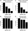
The percentage of absorbance compared with the control under conditions of 2 μM ATP was calculated using the same dataset as shown in Figure 4 at a concentration of 2 μM ATP. The results shown are for unstimulated (A) and EGF-stimulated (C) deletion mutant EGFR and unstimulated (B) and EGF-stimulated (D) wild-type EGFR in response to ATP with or without gefitinib. Results are representative of at least three independent experiments.

Similar articles

Cited by

References

    1. Arteaga C. L. ErbB-targeted therapeutic approaches in human cancer. Exp. Cell Res. 2003;284:122–130. - PubMed
    1. Traxler P., Furet P., Mett H., Buchdunger E., Meyer T., Lydon N. Design and synthesis of novel tyrosine kinase inhibitors using a pharmacophore model of the ATP-binding site of the EGF-R. J. Pharm. Belg. 1997;52:88–96. - PubMed
    1. Shepherd F. A., Rodrigues Pereira J., Ciuleanu T., Tan E. H., Hirsh V., Thongprasert S., Campos D., Maoleekoonpiroj S., Smylie M., Martins R., et al. Erlotinib in previously treated non-small-cell lung cancer. N. Engl. J. Med. 2005;353:123–132. - PubMed
    1. Bell D. W., Lynch T. J., Haserlat S. M., Harris P. L., Okimoto R. A., Brannigan B. W., Sgroi D. C., Muir B., Riemenschneider M. J., Iacona R. B., et al. Epidermal growth factor receptor mutations and gene amplification in non-small-cell lung cancer: molecular analysis of the IDEAL/INTACT gefitinib trials. J. Clin. Oncol. 2005;23:8081–8092. - PubMed
    1. Lynch T. J., Bell D. W., Sordella R., Gurubhagavatula S., Okimoto R. A., Brannigan B. W., Harris P. L., Haserlat S. M., Supko J. G., Haluska F. G., et al. Activating mutations in the epidermal growth factor receptor underlying responsiveness of non-small-cell lung cancer to gefitinib. N. Engl. J. Med. 2004;350:2129–2139. - PubMed

Publication types