. 2006 Apr 19:3:24.
doi: 10.1186/1742-4690-3-24.

Derivation of normal macrophages from human embryonic stem (hES) cells for applications in HIV gene therapy

Affiliations

Derivation of normal macrophages from human embryonic stem (hES) cells for applications in HIV gene therapy

Joseph S Anderson et al. Retrovirology. .

Abstract

Background: Many novel studies and therapies are possible with the use of human embryonic stem cells (hES cells) and their differentiated cell progeny. The hES cell derived CD34 hematopoietic stem cells can be potentially used for many gene therapy applications. Here we evaluated the capacity of hES cell derived CD34 cells to give rise to normal macrophages as a first step towards using these cells in viral infection studies and in developing novel stem cell based gene therapy strategies for AIDS.

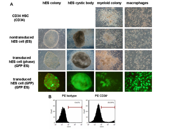
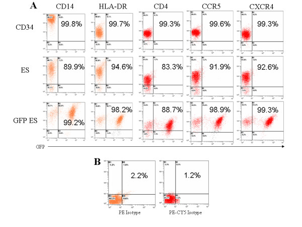
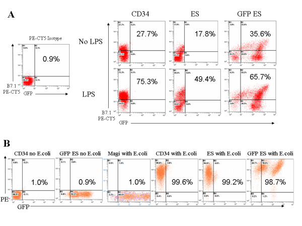
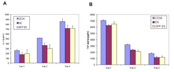
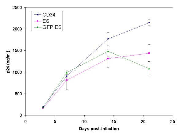
Results: Undifferentiated normal and lentiviral vector transduced hES cells were cultured on S17 mouse bone marrow stromal cell layers to derive CD34 hematopoietic progenitor cells. The differentiated CD34 cells isolated from cystic bodies were further cultured in cytokine media to derive macrophages. Phenotypic and functional analyses were carried out to compare these with that of fetal liver CD34 cell derived macrophages. As assessed by FACS analysis, the hES-CD34 cell derived macrophages displayed characteristic cell surface markers CD14, CD4, CCR5, CXCR4, and HLA-DR suggesting a normal phenotype. Tests evaluating phagocytosis, upregulation of the costimulatory molecule B7.1, and cytokine secretion in response to LPS stimulation showed that these macrophages are also functionally normal. When infected with HIV-1, the differentiated macrophages supported productive viral infection. Lentiviral vector transduced hES cells expressing the transgene GFP were evaluated similarly like above. The transgenic hES cells also gave rise to macrophages with normal phenotypic and functional characteristics indicating no vector mediated adverse effects during differentiation.

Conclusion: Phenotypically normal and functionally competent macrophages could be derived from hES-CD34 cells. Since these cells are susceptible to HIV-1 infection, they provide a uniform source of macrophages for viral infection studies. Based on these results, it is also now feasible to transduce hES-CD34 cells with anti-HIV genes such as inhibitory siRNAs and test their antiviral efficacy in down stream differentiated cells such as macrophages which are among the primary cells that need to be protected against HIV-1 infection. Thus, the potential utility of hES derived CD34 hematopoietic cells for HIV-1 gene therapy can be evaluated.


Figures

Figure 1
Figure 1
Derivation of macrophages from lentiviral vector transduced and normal hES cells. A) Transduced and non-transduced H1 hES cells were cultured on mouse S17 bone marrow stromal cell layers to derive cystic bodies. Cystic body derived CD34 cells were purified by positive selection with antibody conjugated magnetic beads and placed in methocult media to obtain myelomonocytic colonies. Pooled colonies were cultured in liquid cytokine media supplemented with GM-CSF and M-CSF to promote macrophage growth. For comparison, fetal liver derived CD34 cells were cultured similarly to derive macrophages. Representative ES cell colonies, cystic bodies, methocult colonies, and derivative macrophages are shown with GFP expressing cells fluorescing green under UV illumination. B) Representative FACS profile of hES cell derived CD34 cells stained with PE conjugated antibodies. Percent positive CD34 cells are shown with isotype control shown in the left panel.
Figure 2
Figure 2
Phenotypic FACS analysis of hES cell derived macrophages. A) Macrophages derived from transduced and nontransduced hES CD34 and fetal liver CD34 cells were stained with antibodies to CD14, HLA-DR, CD4, CCR5, and CXCR4 and the expression of these surface markers was analyzed by FACS. B) Isotype controls for PE and PE-CY5 antibodies. Percent positive cells are displayed in the plots for each respective cell surface marker staining. Dot plots are representative of triplicate experiments.
Figure 3
Figure 3
Functional analysis of hES cell derived macrophages for B7.1 costimulatory molecule upregulation and phagocytosis of E. coli particles: A) Mature macrophages were stimulated with LPS to determine B7.1 upregulation. Twenty-four hours post-stimulation, macrophages were labeled with a PE-CY5 conjugated anti-B7.1 antibody and analyzed by FACS. B7.1 upregulation data are representative of triplicate experiments. Isotype control is shown in the left panel. B) To assess phagocytic function, E. coli Bioparticles® were added directly to the cultured macrophages. Twenty four hours post-addition, cells were analyzed by FACS. Percent positive cells are displayed in the plots for each experiment. These data are representative of triplicate experiments.
Figure 4
Figure 4
Cytokine IL-1 and TNFα secretion by stimulated hES cell derived macrophages: Macrophages derived from transduced and nontransduced hES and fetal liver CD34 cells were stimulated with 5 μg/ml LPS. On days 1, 2, and 3 post-stimulation, supernatants were collected and assayed by ELISA for (A) IL-1 and (B) TNFα. Experiments were done in triplicate.
Figure 5
Figure 5
hES cell derived macrophages support productive HIV-1 infection: Macrophages derived from transduced and nontransduced hES CD34 and fetal liver CD34 cells were infected with macrophage R5-tropic HIV-1 BaL-1 strain at an m.o.i. of 0.01. Culture supernatants were collected on different days post infection and assayed for viral p24 antigen by ELISA. Data is representative of triplicate experiments.

Similar articles

Cited by

References

    1. Weissman I. Stem Cell Research: Paths to cancer therapies and regenerative medicine. JAMA. 2005;294:1359–1366. doi: 10.1001/jama.294.11.1359. - DOI - PubMed
    1. Lerou PH, Daley GQ. Therapeutic potential of embryonic stem cells. Blood Rev. 2005;19:321–331. doi: 10.1016/j.blre.2005.01.005. - DOI - PubMed
    1. Menendez P, Wang L, Bhatia M. Genetic manipulation of human embryonic stem cells: A system to study early human development andpotential therapeutic applications. Curr Gene Ther. 2005;5:375–385. doi: 10.2174/1566523054546198. - DOI - PubMed
    1. D'Amour KA, Agulnick AD, Eliazar S, Kelly OG, Kroon E, Baetge EE. Efficient differentiation of human embryonic stem cells to definitive endoderm. Nat Biotechnol. 2005;23:1534–1541. doi: 10.1038/nbt1163. - DOI - PubMed
    1. Thomson JA, Itskovitz-Eldor J, Shapiro SS, Waknitz MA, Swiergiel JJ, Marshall VS, Jones JM. Embryonic stem cell lines derived from human blastocysts. Science. 1998;282:1145–1147. doi: 10.1126/science.282.5391.1145. - DOI - PubMed

Publication types