. 2006 Apr 19:6:97.
doi: 10.1186/1471-2407-6-97.

Nuclear FABP7 immunoreactivity is preferentially expressed in infiltrative glioma and is associated with poor prognosis in EGFR-overexpressing glioblastoma

Affiliations

Nuclear FABP7 immunoreactivity is preferentially expressed in infiltrative glioma and is associated with poor prognosis in EGFR-overexpressing glioblastoma

Yu Liang et al. BMC Cancer. .

Abstract

Background: We previously identified brain type fatty acid-binding protein (FABP7) as a prognostic marker for patients with glioblastoma (GBM). Increased expression of FABP7 is associated with reduced survival. To investigate possible molecular mechanisms underlying this association, we compared the expression and subcellular localization of FABP7 in non-tumor brain tissues with different types of glioma, and examined the expression of FABP7 and epidermal growth factor receptor (EGFR) in GBM tumors.

Methods: Expression of FABP7 in non-tumor brain and glioma specimens was examined using immunohistochemistry, and its correlation to the clinical behavior of the tumors was analyzed. We also analyzed the association between FABP7 and EGFR expression in different sets of GBM specimens using published DNA microarray datasets and semi-quantitative immunohistochemistry. In vitro migration was examined using SF763 glioma cell line.

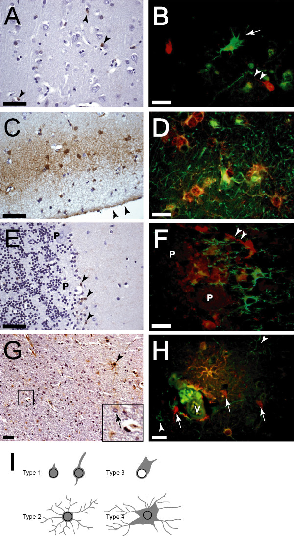
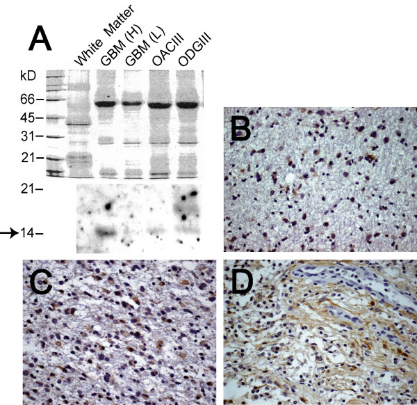
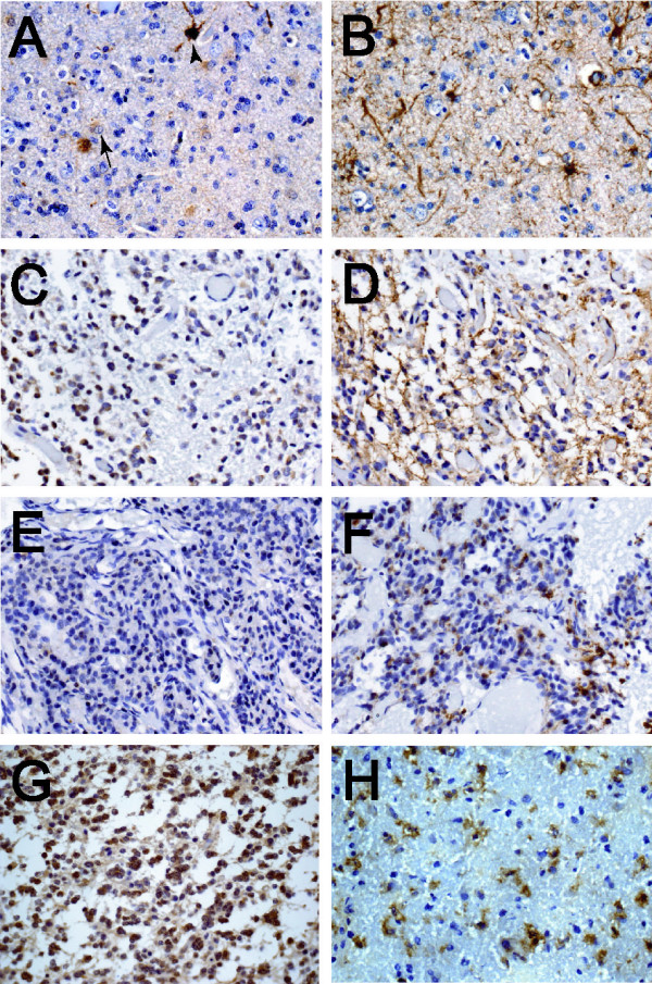
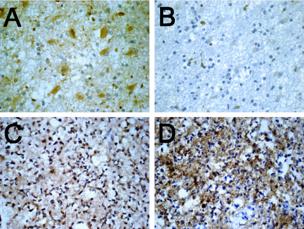
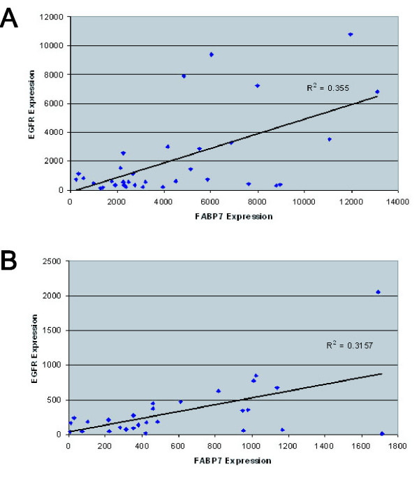
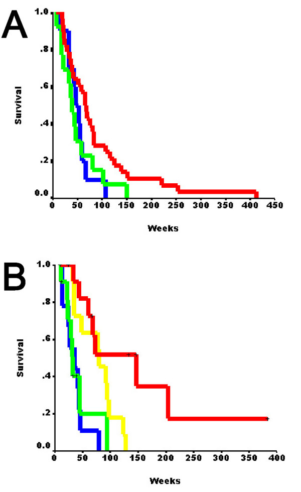
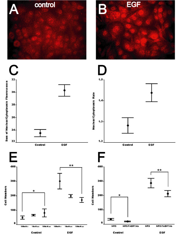
Results: FABP7 was present in a unique population of glia in normal human brain, and its expression was increased in a subset of reactive astrocytes. FABP7 immunoreactivity in grade I pilocytic astrocytoma was predominantly cytoplasmic, whereas nuclear FABP7 was detected in other types of infiltrative glioma. Nuclear, not cytoplasmic, FABP7 immunoreactivity was associated with EGFR overexpression in GBM (N = 61, p = 0.008). Expression of the FABP7 gene in GBM also correlated with the abundance of EGFR mRNA in our previous microarray analyses (N = 34, p = 0.016) and an independent public microarray dataset (N = 28, p = 0.03). Compared to those negative for both markers, nuclear FABP7-positive/EGFR-positive and nuclear FABP7-positive/EGFR-negative GBM tumors demonstrated shortest survival, whereas those only positive for EGFR had intermediate survival. EGFR activation increased nuclear FABP7 immunoreactivity in a glioma cell line in vitro, and inhibition of FABP7 expression suppressed EGF-induced glioma-cell migration. Our data suggested that in EGFR-positive GBM the presence of nuclear FABP7 immunoreactivity increases the risk of poor prognosis

Conclusion: In this study, we identified a possible mechanism as the basis of the association between nuclear FABP7 and poor prognosis of GBM. FABP7 expression can be found in all grades of astrocytoma, but neoplastic cells with nuclear FABP7 were only seen in infiltrative types of tumors. Nuclear FABP7 may be induced by EGFR activation to promote migration of GBM tumor cells. Positive nuclear FABP7 and EGFR overexpression correlated with short survival in EGFR-positive GBM patients. Therefore, nuclear FABP7 immunoreactivity could be used to monitor the progression of EGFR-overexpressed GBM.


Figures

Figure 1
Figure 1
Several groups of glial cells in adult normal and gliotic brains expressed FABP7. A, scattered FABP7-positive Type 1 cells (arrowheads) were detected in normal cerebral cortex. Nuclei of these cells were irregular or elongated, and appeared to be larger than those of oligodendrocytes. B, FABP7-positive Type 1 cells usually had only one or two processes (arrowheads), and were distinctive from GFAP-expressing astrocytes bearing elaborate processes (arrow). C and D, all FABP7-positive Type 2 cells localized at the subpial layer had numerous processes and expressed GFAP. Arrowheads in C indicate pia. Some GFAP+/FABP7- astrocytes could be seen. E, scattered FABP7-positive Type 1 cells were detected in normal cerebellum (arrowheads), whereas granule cells and Purkinje cells (P) were FABP7 negative. F, FABP7 and GFAP staining identified two distinctive populations of astrocytes. Arrowheads indicate the process of a FABP7-positive Type 1 cell. G, the number of FABP7-positive cells was increased in gliotic tissues, and Types 3 (arrow in inset) and 4 (arrowhead) cells appeared. H, in a region of perivascular gliosis, several GFAP+/FABP+ reactive astrocytes (Type 4) were seen. There were also FABP7-negative reactive astrocytes (arrowheads) and Type 1 cells (arrows) in this region. Strong immunofluorescence inside the vessel (V) was autofluorescence. I is a schematic summary of the morphology of the four types of glial cells identified in normal and gliotic adult brains. Gray color indicates FABP7 immunoreactivity. Note that Type 3 cells have gemistocytic characteristics and usually do not have nuclear FABP7. Bars in A,C,E,G, 50μm; bars in B, D, F, H, 20μm, and red and green fluorescence represents FABP7 and GFAP, respectively.
Figure 2
Figure 2
FABP7 was detected in all grades of astrocytomas. A, lower, expression of FABP7 was analyzed Western blotting using lysate from normal white matter, two GBM specimens (H and L indicate high and low, respectively (high and low FABP7 expression based on the results of the previous microarray study [9]), one oligoastrocytoma grade III (OACIII), and one oligodendroglioma grade III (ODGIII); upper, the protein loading was demonstrated by Coomassie Blue staining. The arrow indicates the size of the FABP7 core protein. The 14 kD band in GBM (L) appeared after longer exposure of the film. The 18 kD band that appeared in the ODGIII lysate may not be non-specific, since several immunoreactive bands in that region could also be detected in lysate from a panel of glioma cell lines (data not shown). Representative photomicrographs of immunohistochemistry of FABP7 in grade II astrocytoma (B), grade III anaplastic astrocytoma (C), and grade I pilocytic astrocytoma (D) demonstrated distinctive FABP7 immunoreactivity in the cytoplasm and cell processes in pilocytic astrocytomas as opposed to the nuclear and cytoplasmic staining in grades II and III astrocytomas. FABP7-positive nuclei that were occasionally detected resembled the Type 1 cells seen in normal brain. The scale of the photomicrographs is the same as in Figure 1 A.
Figure 3
Figure 3
FABP7 is expressed in GFAP-positive cells in ODG and OAC. In grade III ODG, FABP7 (A) was expressed only in a subset of reactive astrocytes resembling Types 3 (arrow) and 4 (arrowhead) cells when compared to numerous GFAP-positive cells in an adjacent section (B). FABP7 was expressed in both nuclei and cytoplasm of the microgemistocytes (C) that were GFAP-immunoreactive (D). However, some tumors with microgemistocytes clearly did not express detectable amount of FABP7 (E) compared to GFAP (F). FABP7 was also seen in both nuclei and cytoplasm of tumor cells in some grades II (G) and III (H) OAC, and adjacent sections showed that the same groups of cells were GFAP positive (data not shown). The scale of the photomicrographs is the same as in Figure 1 A.
Figure 4
Figure 4
EGFR expression is associated with nuclear but not cytoplasmic FABP7 immunoreactivity. In one region of a representative GBM specimen, the cytoplasm of neoplastic gemistocytes had pronounced FABP7 immunoreactivity but the nuclei of the same cells were negative (A). In an adjacent section of the same specimen only minimal EGFR staining was seen in the same population of tumor cells (B). In another region of the same specimen, all nuclei of neoplastic astrocytes were marked by FABP7 staining (C), and EGFR expression was also prominent in these cells (D). The scale of photomicrographs is the same as in Figure 1 A.
Figure 5
Figure 5
Scattered plot analyses of the expression levels of FABP7 and EGFR genes. The abundance of mRNA for both genes obtained by microarrays was presented on a log2 base. A strong positive correlation of FABP7 and EGFR expression was seen in a set of 34 GBM specimens (A, p = 0.016), which was validated by a public microarray dataset using 28 GBM specimens (B, p = 0.03).
Figure 6
Figure 6
Kaplan-Meier analysis of the first set of 55 GBM patients based on the new categories (0, N = 28; 1, N = 4; 2, N = 13; 3, N=10) of nuclear FABP7 and EGFR expression (see Methods) showed that dual-negative specimens had significant longer survival time (A). Kaplan-Meier analysis of an independent set of 44 younger GBM patients (0, N = 11; 1, N = 12; 2, N = 11; 3, N=10) showed gradual decrease of survival time based on increased expression of nuclear FABP7 and EGFR (B). Red, category 0; yellow, category 1; green, category 2; blue, category 3.
Figure 7
Figure 7
EGF activation induces nuclear translocation of FABP7 in glioma cells. Immunofluorescent staining of FABP7 in SF763 glioma cells displayed both cytoplasmic and nuclear immunoreactivity (A), whereas FABP7 staining in the nuclei was increased after EGF treatment for 2 days (B). The scale of photomicrographs is the same as in Figure 1A. C, the sum of nuclear and cytoplasmic FABP7 fluorescence in SF763 cells in each of the 10 randomly chosen fields was calculated, and EGF treatment significantly increased total FABP7 fluorescence. D, the ratios of nuclear/cytoplasmic FABP7 in untreated SF763 glioma cells and cells treated with 50 ng/ml of EGF for 1 day were semi-quantitatively measured. For each group, ratios of 20 cells were calculated from each of the 10 randomly chosen fields (p < 0.001). E, control SF763 cells demonstrated no difference in migration in the presence of either FABP7-specific sense or antisense ODNs (*, p = 0.136), whereas migration of EGF-treated SF763 cells was reduced by more than one-third in the presence of antisense ODNs (**, p = 0.004). F, FABP7 polyclonal antibodies also inhibited migration of both control and EGF-treated SF763 cells (*, p=0.003; **, p=0.01). Error bars represented the standard errors of the mean.

References

    1. Medical Research Council Brain Tumor Working Party Randomized trial of procarbazine, lomustine, and vincristine in the adjuvant treatment of high-grade astrocytoma: a Medical Research Council trial. J Clin Oncol. 2001;19:509–518. - PubMed
    1. Korkolopoulou P, Christodoulou P, Kouzelis K, Hadjiyannakis M, Priftis A, Stamoulis G, Seretis A, Thomas-Tsagli E. MDM2 and p53 expression in gliomas: a multivariate survival analysis including proliferation markers and epidermal growth factor receptor. Br J Cancer. 1997;75:1269–1278. - PMC - PubMed
    1. Etienne MC, Formento JL, Lebrun-Frenay C, Gioanni J, Chatel M, Paquis P, Bernard C, Courdi A, Bensadoun RJ, Pignol JP, Francoual M, Grellier P, Frenay M, Milano G. Epidermal growth factor receptor and labeling index are independent prognostic factors in glial tumor outcome. Clin Cancer Res. 1998;4:2383–2390. - PubMed
    1. Rainov NG, Dobberstein KU, Bahn H, Holzhausen HJ, Lautenschlager C, Heidecke V, Burkert W. Prognostic factors in malignant glioma: influence of the overexpression of oncogene and tumor-suppressor gene products on survival. J Neurooncol. 1997;35:13–28. doi: 10.1023/A:1005841520514. - DOI - PubMed
    1. Waha A, Baumann A, Wolf HK, Fimmers R, Neumann J, Kindermann D, Astrahantseff K, Blumcke I, von Deimling A, Schlegel U. Lack of prognostic relevance of alterations in the epidermal growth factor receptor-transforming growth factor-alpha pathway in human astrocytic gliomas. J Neurosurg. 1996;85:634–641. - PubMed

Publication types

MeSH terms