

Complementation of Saccharomyces cerevisiae coq7 mutants by mitochondrial targeting of the Escherichia coli UbiF polypeptide: two functions of yeast Coq7 polypeptide in coenzyme Q biosynthesis
- PMID: 16624818
- PMCID: PMC3066048
- DOI: 10.1074/jbc.M513267200
Complementation of Saccharomyces cerevisiae coq7 mutants by mitochondrial targeting of the Escherichia coli UbiF polypeptide: two functions of yeast Coq7 polypeptide in coenzyme Q biosynthesis
Abstract
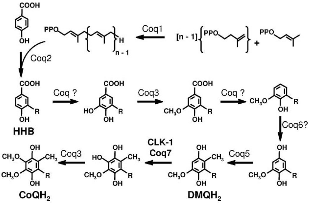
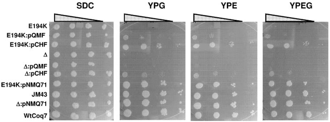
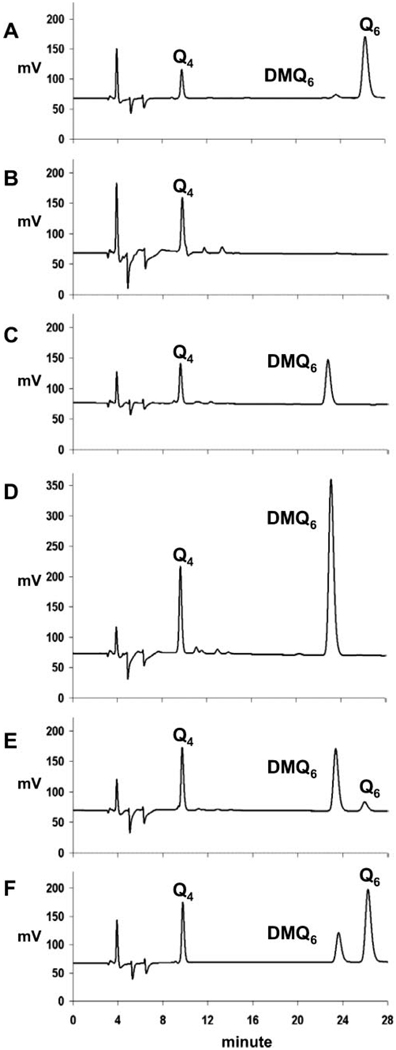
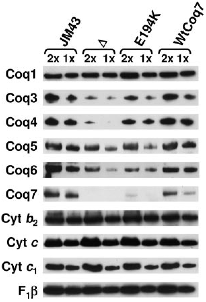
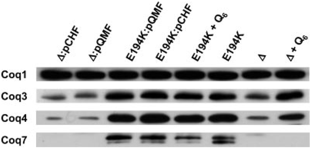
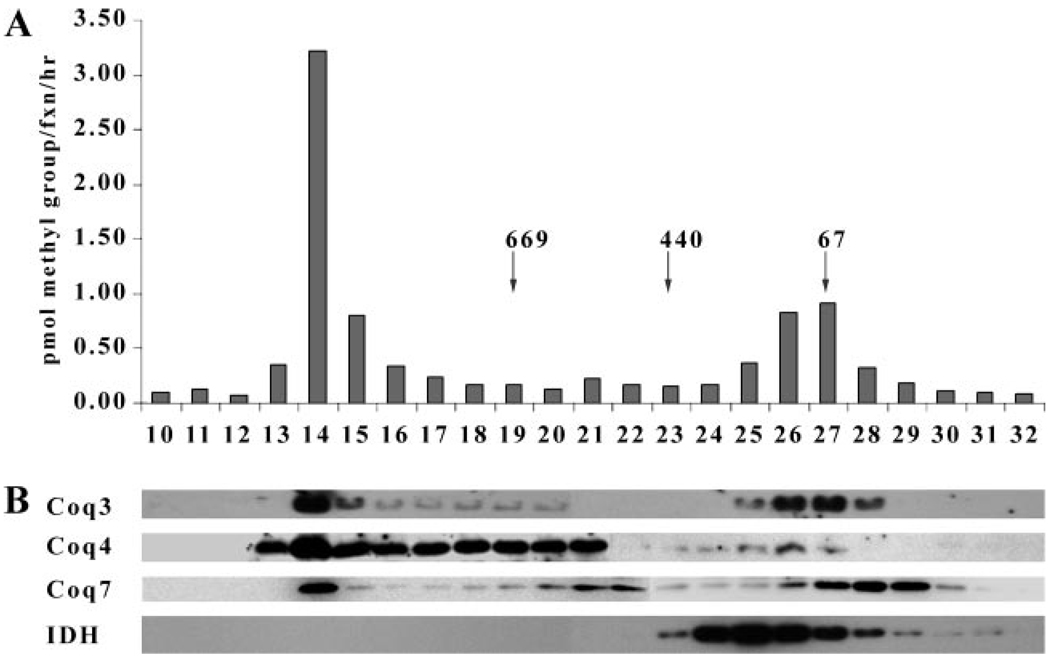
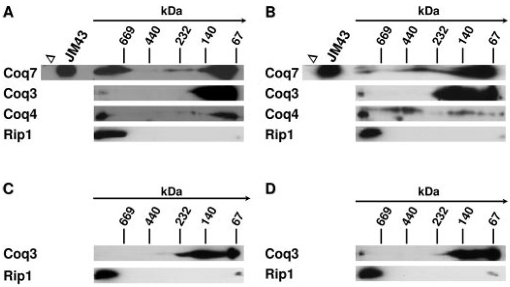
Coenzyme Q (ubiquinone or Q) functions in the respiratory electron transport chain and serves as a lipophilic antioxidant. In the budding yeast Saccharomyces cerevisiae, Q biosynthesis requires nine Coq proteins (Coq1-Coq9). Previous work suggests both an enzymatic activity and a structural role for the yeast Coq7 protein. To define the functional roles of yeast Coq7p we test whether Escherichia coli ubiF can functionally substitute for yeast COQ7. The ubiF gene encodes a flavin-dependent monooxygenase that shares no homology to the Coq7 protein and is required for the final monooxygenase step of Q biosynthesis in E. coli. The ubiF gene expressed at low copy restores growth of a coq7 point mutant (E194K) on medium containing a non-fermentable carbon source, but fails to rescue a coq7 null mutant. However, expression of ubiF from a multicopy vector restores growth and Q synthesis for both mutants, although with a higher efficiency in the point mutant. We attribute the more efficient rescue of the coq7 point mutant to higher steady state levels of the Coq3, Coq4, and Coq6 proteins and to the presence of demethoxyubiquinone, the substrate of UbiF. Coq7p co-migrates with the Coq3 and Coq4 polypeptides as a high molecular mass complex. Here we show that addition of Q to the growth media also stabilizes the Coq3 and Coq4 polypeptides in the coq7 null mutant. The data suggest that Coq7p, and the lipid quinones (demethoxyubiquinone and Q) function to stabilize other Coq polypeptides.
Figures
Similar articles
-
Coenzyme Q supplementation or over-expression of the yeast Coq8 putative kinase stabilizes multi-subunit Coq polypeptide complexes in yeast coq null mutants.Biochim Biophys Acta. 2014 Apr 4;1841(4):630-44. doi: 10.1016/j.bbalip.2013.12.017. Epub 2014 Jan 7. Biochim Biophys Acta. 2014. PMID: 24406904 Free PMC article.
-
The COQ7 gene encodes a protein in saccharomyces cerevisiae necessary for ubiquinone biosynthesis.J Biol Chem. 1996 Feb 9;271(6):2995-3004. doi: 10.1074/jbc.271.6.2995. J Biol Chem. 1996. PMID: 8621692
-
Complementation of Escherichia coli ubiF mutation by Caenorhabditis elegans CLK-1, a product of the longevity gene of the nematode worm.FEBS Lett. 2003 May 22;543(1-3):174-8. doi: 10.1016/s0014-5793(03)00419-8. FEBS Lett. 2003. PMID: 12753928
-
Regulation of coenzyme Q biosynthesis in yeast: a new complex in the block.IUBMB Life. 2014 Feb;66(2):63-70. doi: 10.1002/iub.1243. Epub 2014 Jan 27. IUBMB Life. 2014. PMID: 24470391 Review.
-
Coenzyme Q10 deficiencies: pathways in yeast and humans.Essays Biochem. 2018 Jul 20;62(3):361-376. doi: 10.1042/EBC20170106. Print 2018 Jul 20. Essays Biochem. 2018. PMID: 29980630 Free PMC article. Review.
Cited by
-
Coq6 is responsible for the C4-deamination reaction in coenzyme Q biosynthesis in Saccharomyces cerevisiae.J Biol Chem. 2015 Oct 2;290(40):24140-51. doi: 10.1074/jbc.M115.675744. Epub 2015 Aug 10. J Biol Chem. 2015. PMID: 26260787 Free PMC article.
-
Genetic evidence for an interaction of the UbiG O-methyltransferase with UbiX in Escherichia coli coenzyme Q biosynthesis.J Bacteriol. 2006 Sep;188(17):6435-9. doi: 10.1128/JB.00668-06. J Bacteriol. 2006. PMID: 16923914 Free PMC article.
-
Overexpression of the Coq8 kinase in Saccharomyces cerevisiae coq null mutants allows for accumulation of diagnostic intermediates of the coenzyme Q6 biosynthetic pathway.J Biol Chem. 2012 Jul 6;287(28):23571-81. doi: 10.1074/jbc.M112.360354. Epub 2012 May 16. J Biol Chem. 2012. PMID: 22593570 Free PMC article.
-
Restoring de novo coenzyme Q biosynthesis in Caenorhabditis elegans coq-3 mutants yields profound rescue compared to exogenous coenzyme Q supplementation.Gene. 2012 Sep 10;506(1):106-16. doi: 10.1016/j.gene.2012.06.023. Epub 2012 Jun 23. Gene. 2012. PMID: 22735617 Free PMC article.
-
CoQ10 a super-vitamin: review on application and biosynthesis.3 Biotech. 2018 May;8(5):249. doi: 10.1007/s13205-018-1271-6. Epub 2018 May 9. 3 Biotech. 2018. PMID: 29755918 Free PMC article. Review.
References
-
- Lenaz G, De Santis A. In: Coenzyme Q. Lenaz G, editor. Chichester, UK: John Wiley & Sons; 1985. pp. 165–199.
-
- Turunen M, Olsson J, Dallner G. Biochim. Biophys. Acta. 2004;1660:171–199. - PubMed
-
- Shults CW, Oakes D, Kieburtz K, Beal MF, Haas R, Plumb S, Juncos JL, Nutt J, Shoulson I, Carter J, Kompoliti K, Perlmutter JS, Reich S, Stern M, Watts RL, Kurlan R, Molho E, Harrison M, Lew M. Arch. Neurol. 2002;59:1541–1550. - PubMed
-
- Johnson A, Gin P, Marbois BN, Hsieh EJ, Wu M, Barros MH, Clarke CF, Tzagoloff A. J. Biol. Chem. 2005;280:31397–31404. - PubMed
Publication types
MeSH terms
Substances
Grants and funding
LinkOut - more resources
Full Text Sources
Molecular Biology Databases
