. 2006 Jul;17(7):3009-20.
doi: 10.1091/mbc.e06-01-0080. Epub 2006 Apr 19.

Rtn1p is involved in structuring the cortical endoplasmic reticulum

Affiliations

Rtn1p is involved in structuring the cortical endoplasmic reticulum

Johan-Owen De Craene et al. Mol Biol Cell. 2006 Jul.

Abstract

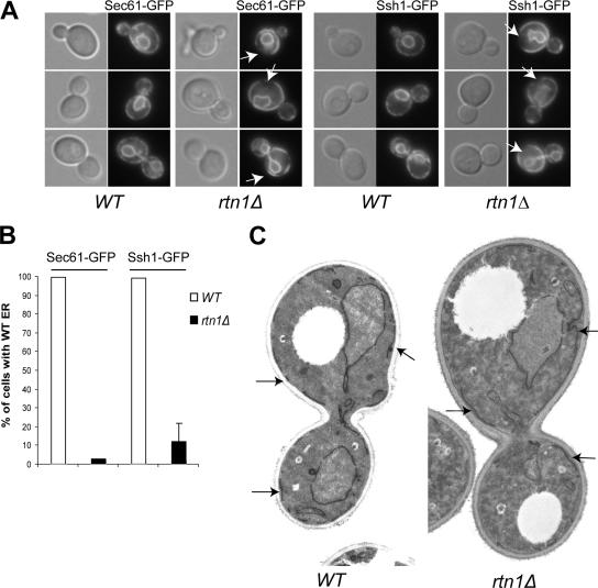
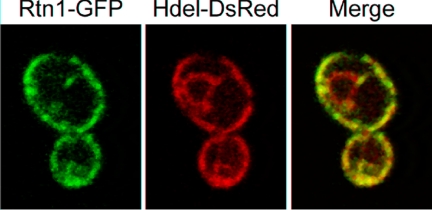
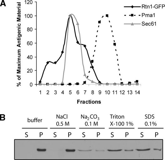
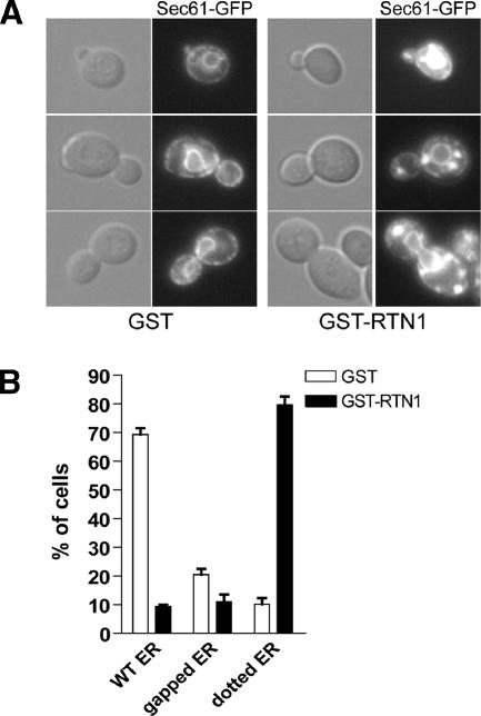
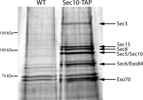
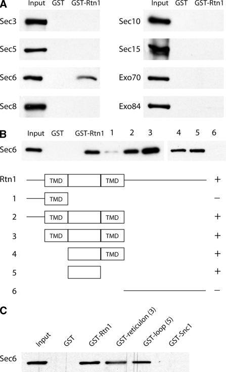
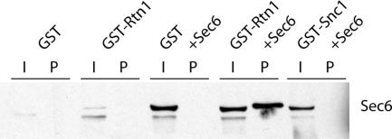
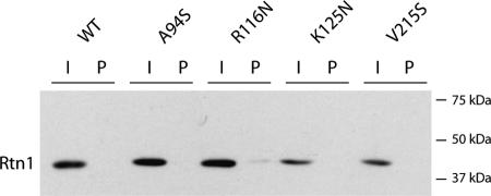
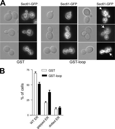
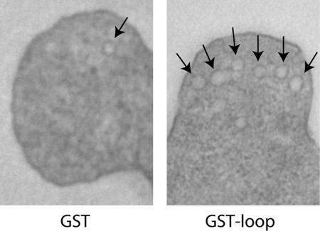
The endoplasmic reticulum (ER) contains both cisternal and reticular elements in one contiguous structure. We identified rtn1Delta in a systematic screen for yeast mutants with altered ER morphology. The ER in rtn1Delta cells is predominantly cisternal rather than reticular, yet the net surface area of ER is not significantly changed. Rtn1-green fluorescent protein (GFP) associates with the reticular ER at the cell cortex and with the tubules that connect the cortical ER to the nuclear envelope, but not with the nuclear envelope itself. Rtn1p overexpression also results in an altered ER structure. Rtn proteins are found on the ER in a wide range of eukaryotes and are defined by two membrane-spanning domains flanking a conserved hydrophilic loop. Our results suggest that Rtn proteins may direct the formation of reticulated ER. We independently identified Rtn1p in a proteomic screen for proteins associated with the exocyst vesicle tethering complex. The conserved hydophilic loop of Rtn1p binds to the exocyst subunit Sec6p. Overexpression of this loop results in a modest accumulation of secretory vesicles, suggesting impaired exocyst function. The interaction of Rtn1p with the exocyst at the bud tip may trigger the formation of a cortical ER network in yeast buds.


Figures

Figure 1.
Figure 1.
Rtn1p is required for the structure of the ER. (A) Wild type (NY2647, NY2649) and rtn1Δ mutant (NY2648, NY2650) cells expressing the ER marker Sec61-GFP or Ssh1-GFP were grown at 25°C in SC medium. The cells were examined using an epifluorescence microscope. Arrows point to cortical regions devoid of fluorescence. (B) Quantification of the altered cortical ER phenotype was done by scoring at least 300 cells with wild-type or altered morphology based on the presence of large gaps in fluorescence at the cell cortex and a more cisternal appearance of the cortical ER in an rtn1Δ mutant. The graph shows the percentage of cells having wild-type ER in each strain. (C) Thin section electron microscopy micrographs of wild-type (NY1210) and rtn1Δ (NY2651) cells grown in YPD and fixed with KMnO4 and embedded in Spurr. Arrows point to ER membrane stretches. Magnification, 4700×.
Figure 2.
Figure 2.
Rtn1-GFP is enriched at the cortical ER. Wild-type cells (NY2658) expressing ER markers Rtn1-GFP and HDEL-DsRed were grown at 25°C in SC medium. The cells were examined using a confocal microscope.
Figure 3.
Figure 3.
Rtn1p is an ER integral membrane protein. (A) Wild-type cells (NY2658) expressing Rtn1-GFP were grown at 25°C in YPD. The lysate was loaded on top of a sucrose density gradient containing EDTA. Rtn1-GFP is represented by the solid black line, the ER marker Sec61p by the solid gray line, and the plasma membrane marker Pma1p by the dashed black line. (B) Wild-type cells (NY2659) expressing Rtn1-3HA were grown at 25°C in SC medium with. Cell lysates were incubated with reagents as indicated and separated into a pellet fraction P and a supernatant fraction S. The fractions were immunoblotted with anti-HA antibodies.
Figure 4.
Figure 4.
Rtn1 overproduction alters ER structure. (A) Wild-type strain expressing the ER marker Sec61-GFP and either GST (NY2660), GST-Rtn1 (NY2661) under the control of the GAL1 were grown at 25°C in SC galactose medium. The cells were examined using an epifluorescence microscope. Arrows point to fluorescence patches. (B) Quantification of the altered cortical ER phenotype was done by scoring at least 300 cells with wild-type or altered morphology based on the presence of large puncta or the presence of large gaps in fluorescence at the cell cortex. The graph shows the percentage of cells having either ER structure.
Figure 5.
Figure 5.
TAP tag isolation of the intact exocyst complex. The exocyst complex was purified from 2 liters of culture using a C-terminal TAP tag on Sec10p. Twenty percent of the sample was run on a 7% polyacrylamide gel and silver stained. The eight exocyst subunits are identified with arrows. Proteins isolated from an untagged strain are shown for comparison.
Figure 6.
Figure 6.
(A) Rtn1p binds to the Sec6p subunit of the exocyst. Soluble recombinant exocyst proteins tagged with 6xHis were incubated with recombinant GST or GST-Rtn1p immobilized on glutathione beads. Approximately 15% of the available Sec6p binds to Rtn1p based in comparison with the 30% input loaded on Western blots. (B) Sec6p binds to the hydrophilic loop of Rtn1p. A deletion series of the Rtn1 protein was constructed and expressed in E. coli. The proteins, immobilized on glutathione beads, were incubated with recombinant 6xHis-Sec6. The hydrophilic loop between the two transmembrane domains (TMD) of Rtn1 was capable of binding 30% of the available Sec6 protein. (C) As a control, GST-Snc1p was included in the binding assay. Again, the Rtn1 loop was sufficient to bind 30% of the available Sec6p, whereas no binding was observed between Sec6p and GST-Snc1p or GST alone.
Figure 7.
Figure 7.
Coprecipitation of Rtn1p and Sec6p when both proteins are overexpressed. The GAL1 promoter was used to overexpress GST, GST-Snc1p, GST-Rtn1p, Sec6p, or combinations of these proteins. GST, GST-Snc1p, or GST-Rtn1p was isolated on glutathione bead, and Sec6p was detected on a Western blot using a Sec6p antibody. The amount of Sec6p is much lower when not driven by the GAL1 promoter, and therefore not as readily detected in samples with endogenous levels. When both Rtn1p and Sec6p are overexpressed in the same strain, ∼0.5% of the available Sec6p (I) is purified (P) with Rtn1p. This band is slightly higher because Sec6p is similar in size to the large amount of Rtn1p isolated, thus causing a slight shift in mobility. No copurification between GST or GST-Snc1 and Sec6 was observed.
Figure 8.
Figure 8.
Orientation of the Rtn1 loop. Three different asparagine-linked glycosylation sites were created in the loop by mutating amino acids 94, 116, and 125 to a serine, asparagine, and asparagines, respectively. A fourth glycosylation site was added near the C terminus of the protein by mutating amino acid 215 to a serine. Soluble protein was isolated and unbound protein (I; 0.25% of total volume) was loaded beside protein (P; 2.5%) that bound to ConA beads. A C-terminal HA tag was used to detect Rtn1p. None of the engineered glycosylation sites caused a shift in mobility, and likewise none of them caused Rtn1p to bind to ConA beads. This negative result suggests these domains are not exposed to the lumen and therefore likely oriented to the cytosol.
Figure 9.
Figure 9.
Rtn1p loop overproduction affects ER structure. (A) Wild-type strain expressing the ER marker Sec61-GFP and either GST (NY2660) or GST-loop (NY2662) under the control of the GAL1 was grown at 25°C in SC galactose medium. The cells were examined using an epifluorescence microscope. Arrows point to fluorescence patches. (B) Quantification of the altered cortical ER phenotype was done by scoring at least 300 cells with wild-type or altered morphology based on the presence of large puncta or the presence of large gaps in fluorescence at the cell cortex. The graph shows the percentage of cells having either ER structure.
Figure 10.
Figure 10.
Rtn1p loop overproduction interferes with secretion. Thin section EM micrographs of wild type transformed either with a 2μ plasmid bearing GST or GST-loop under the control of the GPD1 promoter (NY2678 and NY2679). Cells were grown on SC medium and fixed with glutaraldehyde and stained with OsO4 and embedded in Spurr. Arrows point to vesicles. Magnification, 6500×.

References

    1. Bacon R. A., Salminen A., Ruohola H., Novick P., Ferro-Novick S. The GTP-binding protein Ypt1 is required for transport in vitro: the Golgi apparatus is defective in ypt1 mutants. J. Cell Biol. 1989;109:1015–1022. - PMC - PubMed
    1. Baumann O., Walz B. Endoplasmic reticulum of animal cells and its organization into structural and functional domains. Int. Rev. Cytol. 2001;205:149–214. - PubMed
    1. Bowser R., Muller H., Govindan B., Novick P. Sec8p and Sec15p are components of a plasma membrane-associated 19.5S particle that may function downstream of Sec4p to control exocytosis. J. Cell Biol. 1992;118:1041–1056. - PMC - PubMed
    1. Causton H. C., Ren B., Koh S. S., Harbison C. T., Kanin E., Jennings E. G., Lee T. I., True H. L., Lander E. S., Young R. A. Remodeling of yeast genome expression in response to environmental changes. Mol. Biol. Cell. 2001;12:323–337. - PMC - PubMed
    1. Chen M. S., Huber A. B., van der Haar M. E., Frank M., Schnell L., Spillmann A. A., Christ F., Schwab M. E. Nogo-A is a myelin-associated neurite outgrowth inhibitor and an antigen for mAb IN-1. Nature. 2000;403:434–439. - PubMed

Publication types

MeSH terms

Substances