. 2006 Jul;17(7):2986-95.
doi: 10.1091/mbc.e05-12-1129. Epub 2006 Apr 19.

Forced dimerization of gp130 leads to constitutive STAT3 activation, cytokine-independent growth, and blockade of differentiation of embryonic stem cells

Affiliations

Forced dimerization of gp130 leads to constitutive STAT3 activation, cytokine-independent growth, and blockade of differentiation of embryonic stem cells

Christiane Stuhlmann-Laeisz et al. Mol Biol Cell. 2006 Jul.

Abstract

The mode of activation of glycoprotein 130 kDa (gp130) and the transmission of the activation status through the plasma membrane are incompletely understood. In particular, the molecular function of the three juxtamembrane fibronectin III-like domains of gp130 in signal transmission remains unclear. To ask whether forced dimerization of gp130 is sufficient for receptor activation, we replaced the entire extracellular portion of gp130 with the c-jun leucine zipper region in the chimeric receptor protein L-gp130. On expression in cells, L-gp130 stimulates ligand-independent signal transducer and activator of transcription (STAT) 3 and extracellular signal-regulated kinase 1/2 phosphorylation. gp130 activation could be abrogated by the addition of a competing peptide comprising the leucine zipper region of c-fos. When stably expressed in the interleukin-3-dependent Ba/F3 murine pre-B-cells, these cells showed constitutive STAT3 activation and cytokine-independent growth over several months. Because gp130 stimulation completely suppressed differentiation of murine embryonic stem cells in vitro, we also stably expressed L-gp130 in these cells, which completely blocked their differentiation in the absence of cytokine stimulation and was consistent with high constitutive expression levels of the stem cell factor OCT-4. Thus, L-gp130 can be used in vitro and in vivo to mimic constitutive and ligand-independent activation of gp130 and STAT3, the latter of which is frequently observed in neoplastic diseases.


Figures
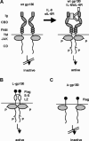
Figure 1.
Figure 1.
Schematic membrane organization and idealized activation of gp130 wild-type and gp130 hybrid receptors. (A) Scheme of the gp130 receptor (wt-gp130) with the extracellular domain comprising Ig-like domain (Ig), cytokine binding domain (CBD), three fibronectin type III-like domains (FNIII), transmembrane domain (TM), and cytoplasmic domain (CD), constitutively associated with JAK. Dimerization is induced after binding of IL-6/sIL-6R. (B) gp130 was truncated 15 amino acids above the transmembrane domain and replaced by the leucine zipper (L) region of the human c-jun gene and stabilized by an additional disulfide bridge. A FLAG-tag (Flag) was inserted between signal peptide and leucine zipper. (C) The extracellular domain of gp130 was removed and only a FLAG-tag was added (Δ-gp130).
Figure 2.
Figure 2.
Functionality of gp130 mutants in transiently transfected COS-7. (A) To demonstrate expression of gp130 and mutants thereof, COS-7 cells were transiently transfected with 5 μg of wt-gp130, L-gp130, and Δ-gp130 cDNAs. Forty-eight hours after transfection, cells were metabolically labeled with [35S]methionine/ cysteine. Cells were lysed, and proteins were precipitated with a gp130-specific antibody. Immune complexes precipitated with protein A-Sepharose were separated by SDS-PAGE and visualized by fluorography. Electrophoretic mobilities of molecular mass marker proteins are indicated on the left. (B) Surface biotinylation of COS-7 cells transfected with Δ-gp130, L-gp130, or wt gp130. (C) To probe for cytokine-independent gp130 phosphorylation, COS-7 cells were mock transfected or transiently transfected with 5 μg of the L-gp130 or Δ-gp130 expression plasmids and lysed 48 h after transfection. Lysates were incubated with a phospho-tyrosine specific antibody (4G10). Precipitated proteins were separated by SDS-PAGE and blotted onto a PVDF membrane. Proteins were detected with a FLAG-specific antibody and visualized by ECL detection.
Figure 3.
Figure 3.
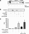
STAT3-activation by gp130 mutants in transiently transfected Cos-7 and HepG2-cells. (A) To analyze STAT3 phosphorylation, transiently transfected HepG2 cells expressing the chimeric receptors and mock-transfected cells were serum starved for 48 h after transfection. Cells were stimulated (+) with 50 ng/ml IL-6 for 10 min or left untreated (−). Lysates were prepared and proteins were precipitated with a phospho-specific STAT3 antibody. Equal amounts of proteins were separated by SDS-PAGE and blotted onto a PVDF membrane. Proteins were detected with a STAT3-specific antibody and visualized by ECL detection. (B) To probe for binding of activated STAT3 to cognate DNA recognition sequences, HepG2 cells were cotransfected with the indicated plasmid combinations. Hyper-IL-6 (50 ng/ml) activated or untreated cells were lysed, and extracts were incubated in the presence of a double-strand biotinylated SIEM oligonucleotide. Protein–oligonucleotide complexes were precipitated with streptavidin-agarose beads. The complexes were subjected to SDS-PAGE, and the separated proteins were transferred onto PVDF membranes. Proteins were detected with the anti-phospho-STAT3 antibody and visualized by ECL detection. Specificity of the observed signal was assessed by adding a 30-fold molar excess of a nonbiotinylated SIEM oligonucleotide during incubation. (C) HepG2 cells were transfected with equal amounts of the indicated plasmids along with the reporter gene constructs SIEM-Luc and pRL-Tk. After serum starvation, cells were stimulated with 50 ng/ml IL-6 or were left untreated for 18 h. Cell lysates were prepared as described, and luciferase activity was measured. Luciferase activity of the samples was normalized to the activity of coexpressed Renilla luciferase. Representative luciferase activities (RLUs) of five independent experiments (each performed in duplicate) are presented.
Figure 4.
Figure 4.
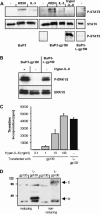
Functionality of gp130 mutants in stable transfected Ba/F3 cell lines. (A) Activation of STAT proteins in Ba/F3 cells: to assay for cellular STAT3/STAT5 activation, equal numbers of Ba/F3, Ba/F3-gp130, and Ba/F3-L-gp130 cells after 4-h serum starvation were stimulated for 10 min with 10% conditioned WEHI-medium (IL-3), 10 ng/ml Hyper-IL-6, or were left untreated. (B) Activation of ERK1/2 proteins in Ba/F3 cells: to assay for cellular ERK1/2 activation, equal numbers of Ba/F3-gp130 and Ba/F3-L-gp130 cells after 4 h serum starvation were stimulated for 10 min with 10 ng/ml Hyper-IL-6 or were left untreated. (C) Equal numbers of stably transfected Ba/F3-L-gp130 cells were cultured for 3 d in the presence or the absence of Hyper-IL-6. Proliferation was measured by pulsing the cells after 68 h with [3H]thymidine for 4 h. Cells were harvested and incorporated radioactivity was measured by scintillation counting. Bioassays were performed with each value being determined in triplicate. (D) Equal numbers of stably transfected Ba/F3-L-gp130 cells and nontransfected Ba/F3 cells were lysed with reducing or nonreducing sample buffer. Equal amounts of proteins were separated by SDS-PAGE and blotted onto a PVDF membrane. Proteins were detected with a anti-FLAG-tag–specific antibody and visualized by ECL detection.
Figure 5.
Figure 5.
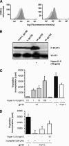
Proliferation of BaF/3-gp130-Δcys-L-gp130 can be inhibited by competitor Fos-peptides. (A) FACS-analysis: the cell lines were labeled as described in Materials and Methods. Cell-associated fluorescence was detected by BD FACSCanto (BD Biosciences). Dark gray curves, control cells Ba/F3-gp130; light gray curves, Ba/F3-gp130 cell lines expressing L-gp130 or Δcys-L-gp130. (B) Activation of STAT proteins in BAF/3 cells: to assay for cellular STAT3 activation, equal numbers of Ba/F3-gp130, Ba/F3-gp130-L-gp130, and Ba/F3-gp130-Δcys-L-gp130 cells after 4-h serum starvation were stimulated for 10 min with 10 ng/ml Hyper-IL-6 (+) or were left untreated (−). Subsequently, cells were lysed and separated by SDS-PAGE. Proteins were transferred onto PVDF membranes. Proteins were detected with a P-STAT3 and a STAT3-specific antibody and visualized by ECL detection. (C) Equal amounts of stable transfected Ba/F3-gp130-L-gp130, Ba/F3-gp130-Δcys-L-gp130, and Ba/F3-gp130 cells were cultured for 3 d in the presence or absence of Hyper-IL-6. Proliferation was measured by pulsing the cells after 68 h with [3H]thymidine for 4 h. Cells were harvested and incorporated radioactivity was measured by scintillation counting. Bioassays were performed with each value being determined in triplicate. (D) Equal amounts of stable transfected Ba/F3-gp130-L-gp130, Ba/F3-gp130-Δcys-L-gp130, and Ba/F3-gp130 cells were cultured for 3 d in the presence of competitor fos peptide. Proliferation was measured by pulsing the cells after 68 h with [3H]thymidine for 4 h. Cells were harvested and incorporated radioactivity was measured by scintillation counting. Bioassays were performed with each value being determined in triplicate.
Figure 6.
Figure 6.
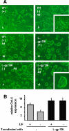
Control of cellular differentiation of embryonic stem cells by L-gp130. (A) Morphological appearance of ES cells expressing L-gp130. Clonally derived ES cell lines expressing L-gp130 (L-gp130) or the empty pEF-BOS expression plasmid (Wt) were routinely maintained in ES cell medium supplemented with 1000 U/ml LIF (+) (i and iv). A single cell suspension of murine Wt ES cells was cultured for 4 d in the presence (iii) or absence (ii) of recombinant LIF, which induces the characteristic change of morphology from densely packed ball-like structure of pluripotent cells to differentiated cells of flattened, fibroblast-like morphology. By contrast, L-gp130–expressing ES cell lines retain their undifferentiated morphology irrespective of the absence (v) or presence (vi) of LIF in the culture medium. (B) Quantitative RT-PCR for Oct-4 expression in murine Wt ES cells or cells expressing L-gp130 (L-gp130) grown for 4 d in the absence (−) or presence of 1000 units/ml LIF (+). RNA extracted from cultures of cells from two independently derived cell lines for each genotype was prepared and analyzed for Oct-4 expression as described previously. Results are expressed as the ratio of Oct-4-18S expression, which served as a control for the amount of RNA analyzed, and data are presented from replicate analysis as the mean ± SD.

References

    1. Auguste P., Guillet C., Fourcin M., Olivier C., Veziers J., Pouplard Barthelaix A., Gascan H. Signaling of type II oncostatin M receptor. J. Biol. Chem. 1997;272:15760–15764. - PubMed
    1. Beadling C., Ng J., Babbage J. W., Cantrell D. A. Interleukin-2 activation of STAT5 requires the convergent action of tyrosine kinases and a serine/threonine kinase pathway distinct from the Raf1/ERK2 MAP kinase pathway. EMBO J. 1996;15:1902–1913. - PMC - PubMed
    1. Behncken S. N., Billestrup N., Brown R., Amstrup J., Conway-Campbell B., Waters M. J. Growth hormone (GH)-independent dimerization of GH receptor by a leucine zipper results in constitutive activation. J. Biol. Chem. 2000;275:17000–17007. - PubMed
    1. Boulanger M. J., Chow D. C., Brevnova E. E., Garcia K. C. Hexameric structure and assembly of the interleukin-6/IL-6 alpha-receptor/gp130 complex. Science. 2003;300:2101–2104. - PubMed
    1. Bromberg J. Stat proteins and oncogenesis. J. Clin. Investig. 2002;109:1139–1142. - PMC - PubMed

Publication types

MeSH terms