. 2006 May 2;103(18):6817-22.
doi: 10.1073/pnas.0602109103. Epub 2006 Apr 19.

Site-specific proteolysis of the transcriptional coactivator HCF-1 can regulate its interaction with protein cofactors

Affiliations

Site-specific proteolysis of the transcriptional coactivator HCF-1 can regulate its interaction with protein cofactors

Jodi L Vogel et al. Proc Natl Acad Sci U S A. .

Abstract

Limited proteolytic processing is an important transcriptional regulatory mechanism. In various contexts, proteolysis controls the cytoplasmic-to-nuclear transport of important transcription factors or removes domains to produce factors with altered activities. The transcriptional coactivator host cell factor-1 (HCF-1) is proteolytically processed within a unique domain consisting of 20-aa reiterations. Site-specific cleavage within one or more repeats generates a family of amino- and carboxyl-terminal subunits that remain tightly associated. However, the consequences of HCF-1 processing have been undefined. In this study, it was determined that the HCF-1-processing domain interacts with several proteins including the transcriptional coactivator/corepressor four-and-a-half LIM domain-2 (FHL2). Analysis of this interaction has uncovered specificity with both sequence and context determinants within the reiterations of this processing domain. In cells, FHL2 interacts exclusively with the nonprocessed coactivator and costimulates transcription of an HCF-1-dependent target gene. The functional interaction of HCF-1 with FHL2 supports a model in which site-specific proteolysis regulates the interaction of HCF-1 with protein partners and thus can modulate the activity of this coactivator. This paradigm expands the biological significance of limited proteolytic processing as a regulatory mechanism in gene transcription.


Conflict of interest statement

Conflict of interest statement: No conflicts declared.

Figures
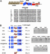
Fig. 1.
Fig. 1.
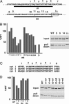
HCF-1–FHL2 interaction mediated by the central region of the PPD. (A) The HCF-1 PPD is shown relative to the amino-terminal (kelch and basic) and the carboxyl-terminal (TA, transactivation; FN3, fibronectin type III; NL, nuclear localization signal) domains. An alignment of the 20 amino acid consensus repeats (blue ovals) and divergent repeats (red ovals) is shown. (B) The HCF-1 PPD contains consensus repeats (blue ovals, 1–2–3, 4–5, and 6), divergent repeats (red ovals, d1–d2 and d3), and an LXXLL motif (yellow circles, L). GST or GST-FHL2 fusion proteins (3 μg) were incubated with the illustrated PPD proteins (10 fmol). The amount of protein bound is expressed as a percent of the amount of bound full-length PPD (repeats 1–6). The results shown are the averages of several independent experiments. Each set of lanes represents input (10% of the total PPD protein in each reaction), GST elutate, and GST-FHL2 elutate. p40, nucleolar protein p40/EBP2.
Fig. 2.
Fig. 2.
Interaction specificity encoded within the HCF-1 PPD consensus repeats. GST or GST-FHL2 fusion proteins were incubated with WT or mutant PPD proteins. (A) The numbers above the sequence of d1 and d2 divergent repeats denote the cluster of amino acids that were changed to alanine to generate the mutant PPD proteins. LL indicates the two amino acids in the LXXLL motif (boxed) that were changed to alanine. (B) The amount of bound protein is graphically represented relative to the amount of WT protein bound (100%) and is representative of several independent experiments. Input was 10% of the total input of PPD protein in each reaction. The autoradiogram shows the results of the mutant PPD proteins (5, 6, 14, and LL) that were impaired in the interaction with FHL2 relative to the WT protein. (C) The consensus repeats 1, 2, and 3 are aligned, and the numbers denote the clusters of amino acids that were changed. (D) The graph and gel show the results of those PPD proteins having mutations in equivalent positions (7 and 8) of repeat 1, 2, or 3.
Fig. 3.
Fig. 3.
Preferential interaction of FHL2 with the 220-kDa HCF-1 precursor. (A) The noncleavable HCF-1 PPD (HCF-1nc) is illustrated with the cleavage site in each repeat (E) mutated to (A). CV-1 cells were transfected with V5-tagged WT or HCF-1nc proteins. Cell lysates were probed using anti-V5 antiserum. (B) CV-1 cells were cotransfected with FLAG-tagged FHL2 or VP16 and the V5-tagged WT HCF-1 or HCF-1nc proteins. FHL2 or VP16 were immunoprecipitated using anti-FLAG serum and probed with anti-V5 and anti-FLAG seras. wt, wild-type; nc, noncleavable; Extract, cell lysate; IP, immunoprecipitate. (C) CV-1 cells were cotransfected with Gal4DB-FHL2 fusion, a Gal4-luciferase reporter, and increasing amounts (80–480 ng) of the WT HCF-1, HCF-1nc, or the control vector. The fold activation is the luciferase activity of the cotransfected cells divided by the basal level and is representative of several independent experiments. (D) Equivalent amounts of extracts of CV-1 cells cotransfected with increasing amounts of WT or HCF-1nc were probed using anti-V5 antisera and anti-β-tubulin (Tb) sera. The amount of full-length HCF-1 protein (FL) and HCF-1 subunit forms were quantitated by using a Kodak Image Station 4400 and normalized to the amount of the β-tubulin internal control.
Fig. 4.
Fig. 4.
FHL2 interacts with the HSV IE accessory factors and costimulates IE gene expression. (A) A typical HSV IE gene (IE-4) is illustrated showing the binding sites for the enhancer core (O-V-H), GABP, and Sp1 that are present in each of the IE gene promoters. (B) GST or GST-FHL2 fusion protein was incubated with 2 fmol of GABPα, GABPβ, Sp1, or the control proteins luciferase (Luc) or p40. Bound proteins were quantitated relative to the amount of protein added to each reaction. Input lanes contained 10% of the labeled protein in each reaction. I, input; G, Gst; F, Gst-FHL2. (C) CV-1 cells were cotransfected with reporters containing the promoters of HSV IE-4, IE-0, EKL-1, or Sp1 (150 ng) and increasing amounts of FHL2, FHL1, or the control vector as indicated. The fold activations represent the luciferase activities of the cotransfected cells divided by the basal levels. The fold activation averages from several independent experiments are graphed (SD ≤ 0.1-fold).
Fig. 5.
Fig. 5.
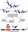
Proteolytic regulation of HCF-1 interactions and coactivation potential. Shown is a model in which processing at HCF-1 reiterations determines the ability of HCF-1 to interact with protein partners (FHL2), thus modulating the HCF-1 coactivation potential. The determinants involved in the interaction of FHL2 with the HCF-1 PPD are indicated (arrows). On the right side of the figure, site-specific cleavage generates an HCF-1 molecule that retains the high-affinity determinants for binding FHL2. The product of this cleavage may recruit FHL2, resulting in an enhanced HCF-1-dependent transcriptional coactivation of a target gene via factors such as GABP and Sp1. Progressive processing in the cell nucleus may ultimately result in destabilization of the HCF-1–FHL2 complex and down-regulation of the coactivation potential. Conversely, the processing shown on the left side of the figure generates an HCF-1 molecule that would have a low affinity for FHL2, resulting in a reduced level of HCF-1-dependent transcriptional coactivation of the target gene.

Similar articles

Cited by

References

    1. Aza-Blanc P., Ramirez-Weber F. A., Laget M. P., Schwartz C., Kornberg T. B. Cell. 1997;89:1043–1053. - PubMed
    1. Brown M. S., Goldstein J. L. Cell. 1997;89:331–340. - PubMed
    1. Fan C. M., Maniatis T. Nature. 1991;354:395–398. - PubMed
    1. Fortini M. E. Curr. Opin. Cell Biol. 2001;13:627–634. - PubMed
    1. Haze K., Yoshida H., Yanagi H., Yura T., Mori K. Mol. Biol. Cell. 1999;10:3787–3799. - PMC - PubMed

Publication types

MeSH terms