. 2006 Jul;173(3):1197-206.
doi: 10.1534/genetics.106.055392. Epub 2006 Apr 19.

Saccharomyces cerevisiae donor preference during mating-type switching is dependent on chromosome architecture and organization

Affiliations

Saccharomyces cerevisiae donor preference during mating-type switching is dependent on chromosome architecture and organization

Eric Coïc et al. Genetics. 2006 Jul.

Abstract

Saccharomyces mating-type (MAT) switching occurs by gene conversion using one of two donors, HMLalpha and HMRa, located near the ends of the same chromosome. MATa cells preferentially choose HMLalpha, a decision that depends on the recombination enhancer (RE) that controls recombination along the left arm of chromosome III (III-L). When RE is inactive, the two chromosome arms constitute separate domains inaccessible to each other; thus HMRa, located on the same arm as MAT, becomes the default donor. Activation of RE increases HMLalpha usage, even when RE is moved 50 kb closer to the centromere. If MAT is inserted into the same domain as HML, RE plays little or no role in activating HML, thus ruling out any role for RE in remodeling the silent chromatin of HML in regulating donor preference. When the donors MAT and RE are moved to chromosome V, RE increases HML usage, but the inaccessibility of HML without RE apparently depends on other chromosome III-specific sequences. Similar conclusions were reached when RE was placed adjacent to leu2 or arg4 sequences engaged in spontaneous recombination. We propose that RE's targets are anchor sites that tether chromosome III-L in MATalpha cells thus reducing its mobility in the nucleus.


Figures

F<sc>igure</sc> 1.—
Figure 1.—
HMLα is the preferred donor when MATa is located on the left arm in RE+ and RE strains. The coordinates of the loci involved in this experiment are indicated in kilobases. HML usage has been quantified at least three times on independent Southern blots. The means and standard errors are shown. (A) The MAT locus has been moved from its natural locus to the HIS4 locus (ECY359). (B) RE has been deleted (Δ) in a strain bearing MATa on the left arm at the HIS4 locus (ECY360). (C) RE has been reinserted near the right donor HMRα-BamHI (ECY362). (D) Representative Southern blots showing the products of MAT switching in strains bearing MAT on the left arm at the HIS4 locus. Genomic DNA digested with the BamHI and NspI restriction enzymes was probed with a MAT-distal DNA fragment. In addition to the parental MATa fragment, the products MATα (from HMLα) and MATα-BamHI (from HMRα-BamHI) are shown.
F<sc>igure</sc> 2.—
Figure 2.—
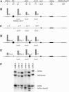
Moving RE to another location on the left arm of chromosome III does not impair its function. (A) Physical map of chromosome III showing the loci involved in this experiment and their position in kilobases. This map is not to scale and the dotted lines represent interruptions in the sequence of the chromosome. (B) Usage of HMLα or of HMRα-LEU2 inserted at different positions on the left arm of chromosome III (41 kb, HIS4, or LEU2) in strains deleted for HMLα (Wu et al. 1996). The donors are represented by shaded horizontal bars. The vertical shaded bars represent the left donor usage as the percentage of a population of switched cells, determined on Southern blots. The solid square represents RE sequences. The solid circle represents the centromere. The horizontal solid rectangle represents MATa. (C) Same as B but the cells are deleted for RE (Δ) (Wu et al. 1996). (D) Same as B but an additional RE is inserted at position 74 kb. (E) Same as D but RE at 29 kb has been deleted. HML usage was measured at least three times on independent Southern blots except when HMRα-LEU2 was inserted at LEU2. Means and standard errors are shown. Strains used in these experiments: (B and C) Wu et al. (1996); (D) 12 kb: GFR18, 41 kb: GFR14, 66 kb: GFR15; (E) 12 kb: GFR22, 41 kb: GFR17, 66 kb: GFR24. (F) Representative Southern blot analysis of the product of mating-type switching in strains bearing either HMLα or (in strains deleted for HMLα) the HMRα donor inserted at different positions on the left arm of chromosome III (Wu et al. 1996). HML usage was quantified on Southern blots after digestion of the genomic DNA with the BamHI and StyI restriction enzymes. A MAT-distal probe detects the products MATα and MATα-BamHI, as well as the parental MATa sequence and a second MAT distal fragment. The extra band observed in the GFR17 corresponds to the unrepaired HO-cut fragment in a fraction of the population.
F<sc>igure</sc> 3.—
Figure 3.—
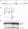
The centromere constitutes a barrier to RE activity. (A) Physical map of chromosome III showing the loci involved in this experiment and their positions in kilobases on the chromosome. (B) Effect of the centromere on RE function. The percentage of HMRα usage (vertical shaded bar) introduced at the LEU2 locus in strains deleted for HML has been measured with RE relocated to three different positions around the centromere: 112.5 kb (ECY139), 115 kb (ECY137), 116.5 kb (ECY138). ECY135 and ECY136 have HML at its normal location and with RE present or deleted, respectively. The solid circle represents the centromere. The solid squares represent RE. (C) To assess the negative effect of the centromere on RE function, the centromere has been moved to the right of RE when at position 116.5 kb (ECY169). Donor usage was quantified at least three times on independent Southern blots; means and standard errors are shown. (D) Representative Southern blot analysis of the product of mating-type switching in strains bearing the RE in the region of the centromere. Genomic DNA was digested with BamHI and HindIII restriction enzymes. Using a Yα fragment as a probe leads to the detection of the products MATα and MATα-BamHI.
F<sc>igure</sc> 4.—
Figure 4.—
Effect of RE on donors located on chromosome V. (A) RE activates poorly a trans-located donor on chromosome V. Donor preference has been measured in strains derived from AN406 in which HMLα has been deleted from chromosome III and integrated near the left telomere of chromosome V at position 9 kb (AN409). The effect of RE on the donor located on chromosome V has been quantified in a strain in which RE has been introduced on chromosome V at position 24 kb (GF26). A representative Southern blot analysis, similar to that carried out in Figure 2, is shown. (B) RE is strongly active on the same chromosome V-located donor when the mating-type switching apparatus is moved entirely on chromosome V. In this experiment, HMLα, MAT, and HMRa have been deleted from chromosome III. HMRα has been reintroduced at position 9 kb on chromosome V, MATa at the ARG5,6 locus, and HMRα-BamHI at the KHS1 locus. HMRα usage has been measured in strains with (ECY429) or without (ECY427) RE at position 24 kb. A representative Southern blot analysis of the product of MAT switching is shown. Genomic DNA was analyzed as in Figure 3. In both experiments, donor usage was measured at least three times on independent Southern blots; means and standard errors are shown.
F<sc>igure</sc> 5.—
Figure 5.—
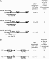
RE depends on chromosome III sequences for its function. (A) Effect of RE on the rates of spontaneous recombination between two leu2 heteroalleles, one located in place of HML on chromosome III and the other integrated at the URA3 locus on chromosome V. Spontaneous rates of recombination have been measured in strains bearing RE at its natural locus (XW485, top), deleted for RE (ECY382, middle), or bearing RE near the leu2 allele located on chromosome V (ECY389, bottom). The proportion of the use of leu2-K as a donor among LEU+ recombinants is also indicated. (B) RE does not induce spontaneous recombination rate outside of chromosome III even when located near a good donor. The spontaneous recombination rate between two arg4 alleles, located at the ARG4 locus on chromosome VIII and at the URA3 locus on chromosome V, is shown in a strain in which RE has been inserted near ARG4 (ECY272) and in the control without RE (ECY269). The proportion of the usage of arg4-Bg as the donor is shown for strain ECY269.

Similar articles

Cited by

References

    1. Amberg, D. C., D. Botstein and E. M. Beasley, 1995. Precise gene disruption in Saccharomyces cerevisiae by double fusion polymerase chain reaction. Yeast 11: 1275–1280. - PubMed
    1. Boeke, J. D., J. Trueheart, G. Natsoulis and G. R. Fink, 1987. 5-Fluoroorotic acid as a selective agent in yeast molecular genetics. Methods Enzymol. 154: 164–175. - PubMed
    1. Bressan, D. A., J. Vazquez and J. E. Haber, 2004. Mating type-dependent constraints on the mobility of the left arm of yeast chromosome III. J. Cell Biol. 164: 361–371. - PMC - PubMed
    1. Chen, D. C., B. C. Yang and T. T. Kuo, 1992. One-step transformation of yeast in stationary phase. Curr. Genet. 21: 83–84. - PubMed
    1. Coïc, E., K. Sun, C. Wu and J. E. Haber, 2006. Cell cycle dependent regulation of Saccharomyces cerevisiae donor preference during mating-type switching by SBF (Swi4/Swi6) and Fkh1. Mol. Cell. Biol. 26: 5470–5480. - PMC - PubMed

Publication types

MeSH terms