. 2006 Jul;173(3):1187-96.
doi: 10.1534/genetics.105.050062. Epub 2006 Apr 19.

Fission yeast Num1p is a cortical factor anchoring dynein and is essential for the horse-tail nuclear movement during meiotic prophase

Affiliations

Fission yeast Num1p is a cortical factor anchoring dynein and is essential for the horse-tail nuclear movement during meiotic prophase

Akira Yamashita et al. Genetics. 2006 Jul.

Abstract

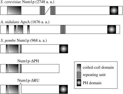
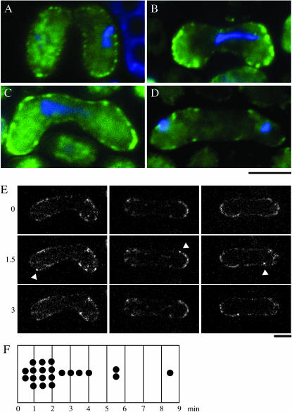
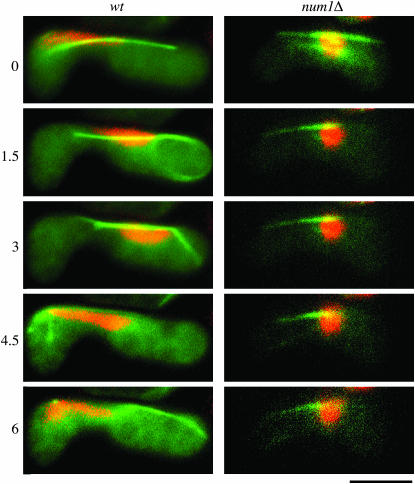
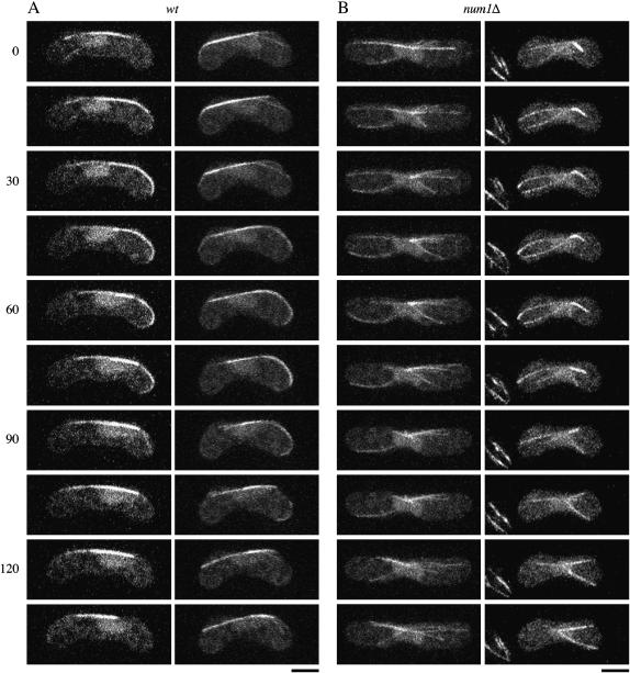
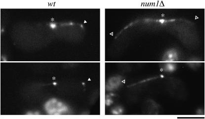
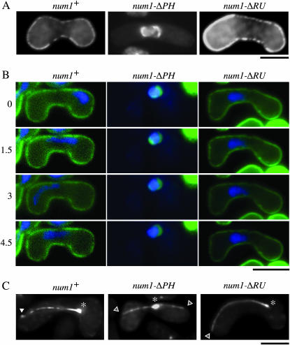
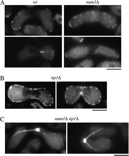
During meiotic prophase in the fission yeast Schizosaccharomyces pombe, the nucleus oscillates between the two ends of a cell. This oscillatory nuclear movement is important to promote accurate pairing of homologous chromosomes and requires cytoplasmic dynein. Dynein accumulates at the points where microtubule plus ends contact the cell cortex and generate a force to drive nuclear oscillation. However, it remains poorly understood how dynein associates with the cell cortex. Here we show that S. pombe Num1p functions as a cortical-anchoring factor for dynein. Num1p is expressed in a meiosis-specific manner and localized to the cell cortex through its C-terminal PH domain. The num1 deletion mutant shows microtubule dynamics comparable to that in the wild type. However, it lacks cortical accumulation of dynein and is defective in the nuclear oscillation as is the case for the dynein mutant. We also show that Num1p can recruit dynein independently of the CLIP-170 homolog Tip1p.


Figures

F<sc>igure</sc> 1.—
Figure 1.—
Schematic of the structure of S. pombe Num1p and its S. cerevisiae and A. nidulans homologs. Num1p-ΔPH lacks the PH domain and Num1p-ΔRU lacks the single conserved repeating unit.
F<sc>igure</sc> 2.—
Figure 2.—
Subcellular localization of Num1p in the course of meiosis. (A–D) Green fluorescence of Num1p-GFP in living cells. Homothallic haploid cells (JV626) carrying the num1-GFP fusion gene were starved for nitrogen to induce conjugation and subsequent meiosis. Nuclear DNA was stained with Hoechst 33342. GFP fluorescence is shown in green, and stained DNA in blue. Bar, 5 μm. Conjugating cells (A), prophase cells (B and C), and a cell at anaphase I (D) are shown. (E) Time-lapse recording of Num1p-GFP. Images were taken at 0.5-min intervals. Z-stacked images of three cells at 1.5-min intervals are shown. Arrowheads indicate GFP fluorescence, which went in and out during a filming period. Bar, 5 μm. (F) The lifetime of a blinking cortical Num1p dot.
F<sc>igure</sc> 3.—
Figure 3.—
Nuclear behavior in wild-type and num1Δ zygotes. Chromosomal DNA in zygotes (JY450 or JV627) was stained with Hoechst 33342 and monitored. The numbers on the left indicate time in minutes. Microtubules were visualized simultaneously by GFP-tagged α-tubulin. Stained DNA is shown in red, and GFP fluorescence in green. Bar, 5 μm.
F<sc>igure</sc> 4.—
Figure 4.—
Microtubule behavior in wild-type and num1Δ zygotes. (A) Wild type (JY450) and (B) num1Δ (JV627) homothallic haploid cells expressing GFP-tagged α-tubulin were starved for nitrogen to induce conjugation and meiosis. Confocal z-step series were recorded at 15-sec intervals and the projection images constructed are shown. The numbers indicate time in seconds. Bar, 5 μm.
F<sc>igure</sc> 5.—
Figure 5.—
Localization of Dhc1p in num1Δ cells. Wild-type (JV897) and num1Δ (JV898) homothallic haploid cells carrying dhc1-GFP were starved for nitrogen and observed under the fluorescence microscope. Asterisks and solid arrowheads indicate GFP fluorescence at SPB and on the cell cortex, respectively. Open arrowheads indicate possible microtubule contact sites on the cell cortex of num1Δ cells, where no accumulation of Dhc1p-GFP was detected. Bar, 5 μm.
F<sc>igure</sc> 6.—
Figure 6.—
Domain analysis of Num1p. (A) num1Δ cells (JV627) were transformed with a plasmid expressing Num1p-GFP, Num1p-ΔPH-GFP, or Num1p-ΔRU-GFP from the nmt1 promoter. Transformants were subjected to nitrogen starvation and observed under the fluorescence microscope. Bar, 5 μm. (B) Nuclear movement during meiotic prophase in the same set of transformants as in A. Chromosomal DNA of each transformant stained with Hoechst 33342 is shown in blue. Fluorescence of GFP-tagged Num1p and its variants is shown in green. The numbers on the left indicate time in minutes. Bar, 5 μm. (C) Localization of Dhc1p in cells expressing Num1p-ΔPH or Num1p-ΔRU. num1Δ homothallic haploid cells (JV656) carrying GFP-dhc1 were transformed with a plasmid expressing Num1p, Num1p-ΔPH, or Num1p-ΔRU from the nmt1 promoter and analyzed as described in Figure 5. Transformants were starved for nitrogen and observed under the fluorescence microscope. Asterisks and solid arrowheads indicate GFP fluorescence at SPB and on the cell cortex, respectively. Open arrowheads indicate possible microtubule contact sites on the cell cortex of num1Δ cells. Bar, 5 μm.
F<sc>igure</sc> 7.—
Figure 7.—
Functional independency of Num1p and Tip1p. (A) Localization of Tip1p during meiotic prophase in wild-type and num1Δ cells. Homothallic haploid wild-type (JV471) and num1Δ cells (JV545) carrying the tip1-GFP fusion gene were starved for nitrogen to induce conjugation and subsequent meiosis and observed under the fluorescence microscope. Bar, 5 μm. (B) Localization of Num1p in tip1Δ cells. Homothallic haploid tip1Δ cells (JV421) carrying the num1-GFP fusion gene were starved for nitrogen to induce conjugation and meiosis and observed under the fluorescence microscope. Bar, 5 μm. (C) Localization of Dhc1p in num1Δ tip1Δ cells. Homothallic haploid num1Δ tip1Δ cells (JV681) carrying GFP-dhc1 were starved for nitrogen and observed under the fluorescence microscope. Bar, 5 μm.

Similar articles

Cited by

References

    1. Adames, N. R., and J. A. Cooper, 2000. Microtubule interactions with the cell cortex causing nuclear movements in Saccharomyces cerevisiae. J. Cell Biol. 149: 863–874. - PMC - PubMed
    1. Bahler, J., J. Q. Wu, M. S. Longtine, N. G. Shah, A. McKenzie III et al., 1998. Heterologous modules for efficient and versatile PCR-based gene targeting in Schizosaccharomyces pombe. Yeast 14: 943–951. - PubMed
    1. Basi, G., E. Schmid and K. Maundrell, 1993. TATA box mutations in the Schizosaccharomyces pombe nmt1 promotor affect transcription efficiency but not the transcription start point or thiamine repressibility. Gene 123: 131–136. - PubMed
    1. Bloom, K., 2001. Nuclear migration: cortical anchors for cytoplasmic dynein. Curr. Biol. 11: R326–R329. - PubMed
    1. Brunner, D., and P. Nurse, 2000. CLIP170-like tip1p spatially organizes microtubular dynamics in fission yeast. Cell 102: 695–704. - PubMed

Publication types

MeSH terms